. 2014 Mar 19;136(11):4382-93.
doi: 10.1021/ja500414a. Epub 2014 Mar 8.

Hairpin DNA sequences bound strongly by bleomycin exhibit enhanced double-strand cleavage

Affiliations

Hairpin DNA sequences bound strongly by bleomycin exhibit enhanced double-strand cleavage

Basab Roy et al. J Am Chem Soc. .

Abstract

Clinically used bleomycin A5 has been employed in a study of double-strand cleavage of a library of 10 hairpin DNAs originally selected on the basis of their strong binding to bleomycin. Each of the DNAs underwent double-strand cleavage at more than one site, and all of the cleavage sites were within, or in close proximity to, an eight-base-pair region of the duplex that had been randomized to create the original library. A total of 31 double-strand cleavage sites were identified on the 10 DNAs, and 14 of these sites were found to represent coupled cleavage sites, that is, events in which one of the two strands was always cleaved first, followed by the associated site on the opposite strand. Most of these coupled sites underwent cleavage by a mechanism described previously by the Povirk laboratory and afforded cleavage patterns entirely analogous to those reported. However, at least one coupled cleavage event was noted that did not conform to the pattern of those described previously. More surprisingly, 17 double-strand cleavages were found not to result from coupled double-strand cleavage, and we posit that these cleavages resulted from a new mechanism not previously described. Enhanced double-strand cleavages at these sites appear to be a consequence of the dynamic nature of the interaction of Fe·BLM A5 with the strongly bound hairpin DNAs.


Figures

Figure 1
Figure 1
Chemical structure of bleomycin A5.
Figure 2
Figure 2
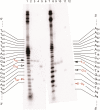
Double-strand cleavage of [5′-32P]-end-labeled (lanes 1–3) and [3′-32P]-end-labeled (lanes 4–6) 64-nucleotide hairpin DNA 8 by Fe(II)·BLM A5. Lane 1, [5′-32P]-end-labeled DNA alone; lane 2, 5 μM Fe(II)·BLM A5; lane 3, 5 μM Fe(II)·BLM A5 + 2 mM n-butylamine; lane 4, 5 μM Fe(II)·BLM A5; lane 5, 5 μM Fe(II)·BLM A5 + 2 mM n-butylamine; lane 6, [3′-32P]-end-labeled DNA alone.
Figure 3
Figure 3
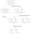
Mechanisms of bleomycin induced double-strand DNA cleavage. Activated Fe·BLM abstracts a H atom from the C-4′position of deoxyribose at the primary site (typically 5′-G-Py-B-3′ of one strand of the hairpin DNA, where B = any nucleobase), producing either an AP (apyrimidinic/apurinic) site (I) or a single-strand break terminating with a 3′-phosphoroglycolate (II). Although the AP site does not undergo further reaction, strand break II is a potential target for a secondary bleomycin cleavage on the opposing strand of the hairpin DNA. The secondary attack of (re)activated bleomycin, by abstracting the C-4′ H atom from the secondary site (5′-B-Pu-C-3′ of the bottom strand of the hairpin DNA) affords either a strand break with a 5′-phosphate/3′phosphoroglycolate or an AP site. The final products are either a double-strand break with 5′-phosphate and 3′-phosphoroglycolate termini (IV) or a strand break at the primary site accompanied by an AP site at the secondary site (III), which upon treatment with mild base (e.g., n-butylamine) produces a double-strand cleavage product (V).
Figure 4
Figure 4
Sequencing gel analysis of Fe(II)·BLM A5-induced double-strand cleavage sites of [3′-32P]-end-labeled (lanes 1–6) and [5′-32P]-end-labeled (lanes 7–12) DNA 8. Each lane (except lanes 1 and 7) corresponds to a numbered cleavage band, shown in Figure 2. Lane 1, Maxam–Gilbert G+A sequencing lane of [3′-32P]-end-labeled DNA 8; lane 2, band 4a; lane 3, band 4b; lane 4, band 5b; lane 5, band 4c; lane 6, band 4d; lane 7, Maxam–Gilbert G+A sequencing lane of [5′-32P]-end-labeled DNA 8; lane 8, band 2a; lane 9, band 2b; lane 10, band 3b; lane 11, band 2c; lane 12, band 2d.
Figure 5
Figure 5
Summary of Fe·bleomycin-induced double-strand cleavage sites on DNAs 2, 8, and 9. Orange bases indicate randomized region of the hairpin DNA library. Arrows of the same shape and color indicate paired cleavages. Black arrows correspond to coupled double-strand cleavage events, whereas red arrows indicate noncoupled double-strand cleavage events, resulting from two independent single-strand cleavages on opposite strands. Nucleotides colored in red indicate primary sites of coupled double-strand DNA cleavage.
Figure 6
Figure 6
Double-strand cleavage of [3′-32P]-end-labeled (lane 2) and [5′-32P]-end-labeled (lane 3) 64-nucleotide hairpin DNA 2 by Fe·BLM A5. Lane 1, [3′-32P]-end-labeled DNA alone; lane 2, 5 μM Fe(II)·BLM A5; lane 3, 5 μM Fe(II)·BLM A5; lane 4, [5′-32P]-end-labeled DNA alone.
Figure 7
Figure 7
Sequencing gel analysis of Fe·bleomycin-induced double-strand cleavage sites of [3′-32P]-end-labeled (lanes 1–7) and [5′-32P]-end-labeled (lanes 8–14) hairpin DNA 2. Each lane (except lanes 1 and 8) corresponds to a numbered cleavage band shown in Figure 6. Lane 1, Maxam–Gilbert G+A sequencing lane of [3′-32P]-end-labeled DNA 2; lane 2, band 2f; lane 3, band 2e; lane 4, band 2d; lane 5, band 2c; lane 6, band 2b; lane 7, band 2a; lane 8, Maxam–Gilbert G+A sequencing lane of [5′-32P]-end-labeled DNA 2; lane 9, band 3f; lane 10, band 3e; lane 11, band 3d; lane 12, band 3c; lane 13, band 3b; lane 14, band 3a.
Figure 8
Figure 8
Analysis of bleomycin-induced double-strand cleavage sites on hairpin DNA 9. (A) Double-strand cleavage of [3′-32P]-end-labeled (lane 2) and [5′-32P]-end-labeled (lane 3) 64-nucleotide hairpin DNA 9 by Fe·bleomycin A5. Lane 1, [3′-32P]-end-labeled DNA alone; lane 2, 5 μM Fe(II)·BLM A5; lane 3, 5 μM Fe(II)·BLM A5; lane 4, [5′-32P]-end-labeled DNA alone. (B) Sequencing gel analysis of Fe·bleomycin-induced double-strand cleavage sites of [3′-32P]-end-labeled (lanes 1–5) and [5′-32P]-end-labeled (lanes 6–10) hairpin DNA 9. Each lane (except lanes 5 and 10) corresponds to a numbered cleavage band, shown in (A). Lane 1, band 2d; lane 2, band 2c; lane 3, band 2b; lane 4, band 2a; lane 5, Maxam–Gilbert G+A sequencing lane of [3′-32P]-end-labeled DNA 9; lane 6, band 3d; lane 7, band 3c; lane 8, band 3b; lane 9, band 3a; lane 10, Maxam–Gilbert G+A sequencing lane of [5′-32P]-end-labeled DNA 9.
Figure 9
Figure 9
Summary of Fe·bleomycin-induced double-strand cleavage sites on hairpin DNAs 110. Orange bases indicate randomized region of the original hairpin DNA library. Arrows of the same shape and color indicate paired cleavages. Black arrows correspond to coupled double-strand cleavage events, whereas red arrows indicate noncoupled double-strand cleavage events, which result from two independent single-strand cleavages on opposite strands. Nucleotides colored in red indicate primary sites of double-strand cleavage.

References

    1. Sausville E. A.; Peisach J.; Horwitz S. B. Biochemistry 1978, 17, 2740. - PubMed
    1. Sausville E. A.; Stein R. W.; Peisach J.; Horwitz S. B. Biochemistry 1978, 17, 2746. - PubMed
    1. Hecht S. M. Acc. Chem. Res. 1986, 19, 383.
    1. Burger R. M. Chem. Rev. 1998, 98, 1153. - PubMed
    1. Povirk L. F. In Molecular Aspects of Anti-Cancer Drug Action; Neidle S., Waring M., Eds.; Macmillan: London, 1983; Vol. 3.

Publication types

LinkOut - more resources