. 2014 Feb 18:11:32.
doi: 10.1186/1742-2094-11-32.

Cerebrospinal fluid detection of interleukin-1β in phase of remission predicts disease progression in multiple sclerosis

Affiliations

Cerebrospinal fluid detection of interleukin-1β in phase of remission predicts disease progression in multiple sclerosis

Silvia Rossi et al. J Neuroinflammation. .

Abstract

Background: Absence of clinical and radiological activity in relapsing-remitting multiple sclerosis (RRMS) is perceived as disease remission. We explored the role of persisting inflammation during remission in disease evolution.

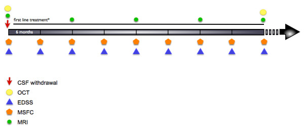
Methods: Cerebrospinal fluid (CSF) levels of interleukin 1β (IL-1β), a major proinflammatory cytokine, were measured in 170 RRMS patients at the time of clinical and radiological remission. These patients were then followed up for at least 4 years, and clinical, magnetic resonance imaging (MRI) and optical coherence tomography (OCT) measures of disease progression were recorded.

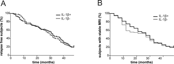
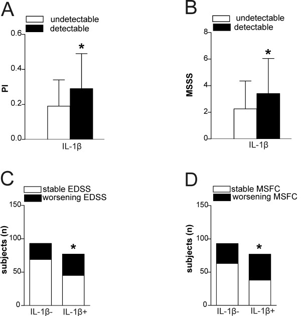
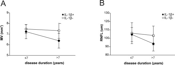
Results: Median follow-up of RRMS patients was 5 years. Detection of CSF IL-1β levels at the time of remission did not predict earlier relapse or new MRI lesion formation. Detection of IL-1β in the CSF was instead associated with higher progression index (PI) and Multiple Sclerosis Severity Scale (MSSS) scores at follow-up, and the number of patients with sustained Expanded Disability Status Scale (EDSS) or Multiple Sclerosis Functional Composite worsening at follow-up was higher in individuals with detectable levels of IL-1β. Patients with undetectable IL-1β in the CSF had significantly lower PI and MSSS scores and a higher probability of having a benign MS phenotype. Furthermore, patients with undetectable CSF levels of IL-1β had less retinal nerve fiber layer thickness and macular volume alterations visualized by OCT compared to patients with detectable IL-1β.

Conclusions: Our results suggest that persistence of a proinflammatory environment in RRMS patients during clinical and radiological remission influences midterm disease progression. Detection of IL-1β in the CSF at the time of remission appears to be a potential negative prognostic factor in RRMS patients.


Figures

Figure 1
Figure 1
Study design. Participants were followed for at least 4 years after cerebrospinal fluid (CSF) collection. Clinical assessment was performed every 6 months, Magnetic resonance imaging (MRI) scans were obtained at least annually. *Second-line treatments were administered to patients who experienced at least two relapses during 1 year of therapy with immunomodulatory agents. EDSS, Expanded Disability Status Scale; MSFC, Multiple Sclerosis Functional Composite; OCT, optical coherence tomography.
Figure 2
Figure 2
Interleukin 1β does not influence disease inflammatory activity in relapsing–remitting multiple sclerosis. (A) and (B) Survival analyses for the time to first clinical relapse (A) and the time to detecting an active magnetic resonance imaging (MRI) scan since diagnosis (B), among participants with detectable or undetectable levels of interleukin 1β (IL-1β) in the cerebrospinal fluid. No significant differences were observed.
Figure 3
Figure 3
Interleukin 1β influences disease progression in relapsing–remitting multiple sclerosis. (A) Progression index (PI) and (B) Multiple Sclerosis Severity Scale (MSSS) scores were higher among participants with detectable interleukin 1β (IL-1β) in the cerebrospinal fluid. Disability progression, measured as a sustained change in score on the Expanded Disability Status Scale (EDSS) (C) or Multiple Sclerosis Functional Composite (MSFC) (D), was higher among participants with detectable IL-1β. *P < 0.05.
Figure 4
Figure 4
Interleukin 1β influences neuronal damage in RR multiple sclerosis. Plots of interaction analysis between interleukin 1β (IL-1β) detection in the cerebrospinal fluid and disease duration, analysis of optical coherence tomography parameters, macular volume (MV) (A) and retinal nerve fiber layer thickness (RNFL) thickness (B). These data confirm the relative preservation of neuronal structures among the participants with undetectable IL-1β, despite disease duration.

References

    1. Rudick RA, Fisher E, Lee JC, Simon J, Jacobs L. Multiple Sclerosis Collaborative Research Group. Use of the brain parenchymal fraction to measure whole brain atrophy in relapsing-remitting MS. Neurology. 1999;53:1698–1704. doi: 10.1212/WNL.53.8.1698. - DOI - PubMed
    1. Rudick RA, Fisher E, Lee JC, Duda JT, Simon J. Brain atrophy in relapsing multiple sclerosis: relationship to relapses, EDSS, and treatment with interferon β-1a. Mult Scler. 2000;6:365–372. - PubMed
    1. Bjartmar C, Kidd G, Mörk S, Rudick R, Trapp BD. Neurological disability correlates with spinal cord axonal loss and reduced N-acetyl aspartate in chronic multiple sclerosis patients. Ann Neurol. 2000;48:893–901. doi: 10.1002/1531-8249(200012)48:6<893::AID-ANA10>3.0.CO;2-B. - DOI - PubMed
    1. Peterson JW, Bö L, Mörk S, Chang A, Trapp BD. Transected neurites, apoptotic neurons, and reduced inflammation in cortical multiple sclerosis lesions. Ann Neurol. 2001;50:389–400. doi: 10.1002/ana.1123. - DOI - PubMed
    1. Meyer R, Weissert R, Diem R, Storch MK, de Graaf KL, Kramer B, Bähr M. Acute neuronal apoptosis in a rat model of multiple sclerosis. J Neurosci. 2001;21:6214–6220. - PMC - PubMed

Publication types

Substances