. 2014 Feb 18;45(1):20.
doi: 10.1186/1297-9716-45-20.

Three viruses of the bovine respiratory disease complex apply different strategies to initiate infection

Affiliations

Three viruses of the bovine respiratory disease complex apply different strategies to initiate infection

Jana Kirchhoff et al. Vet Res. .

Abstract

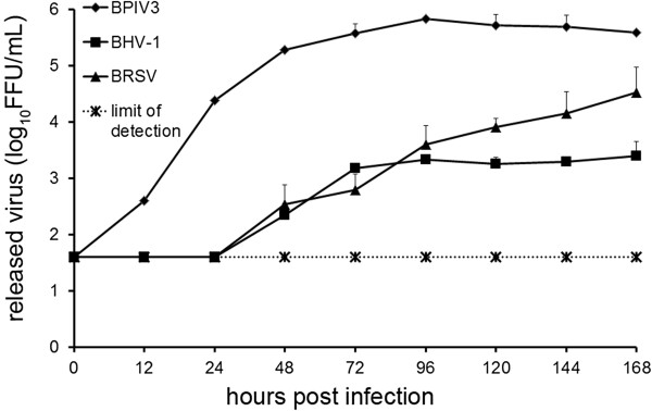
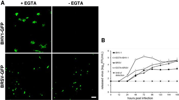
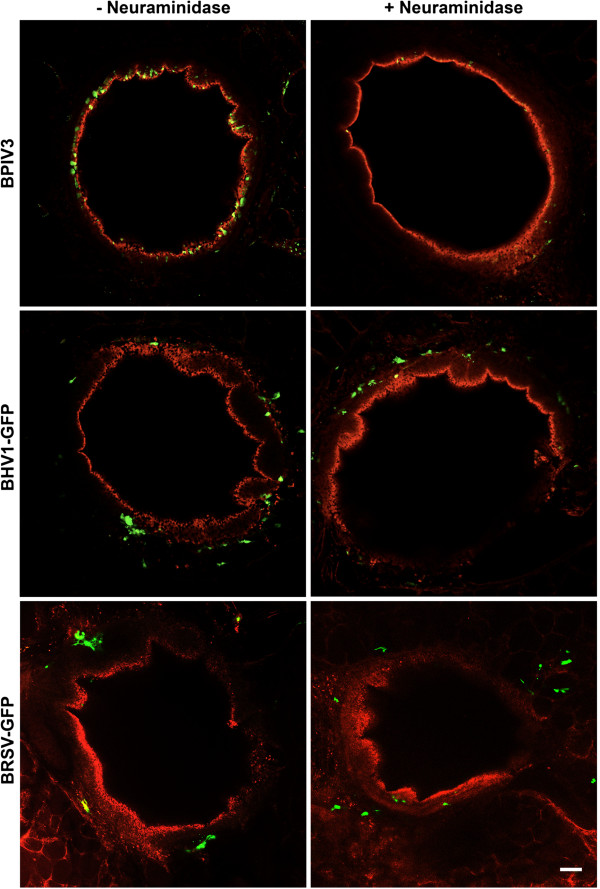
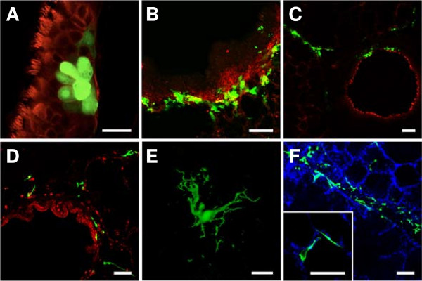
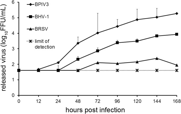
Bovine respiratory disease complex (BRDC) is the major cause of serious respiratory tract infections in calves. The disease is multifactorial, with either stress or reduced immunity allowing several pathogens to emerge. We investigated the susceptibility of bovine airway epithelial cells (BAEC) to infection by the three major viruses associated with the BRDC: bovine respiratory syncytial virus (BRSV), bovine herpesvirus type 1 (BHV-1) and bovine parainfluenza virus type 3 (BPIV3). For this purpose, two culture systems for well-differentiated BAEC were used: the air-liquid interface (ALI) system, where filter-grown BAEC differentiate into a pseudostratified respiratory epithelium and precision-cut lung slices (PCLS) where BAEC are maintained in the original tissue organisation. Comparative infection studies demonstrated that entry and release of BPIV3 occurred specifically via the apical membrane with ciliated cells being the major target cells. By contrast, airway epithelial cells were largely resistant to infection by BHV-1. When the epithelial barrier was abolished by opening tight junctions or by injuring the cell monolayer, BHV-1 infected mainly basal cells. Respiratory epithelial cells were also refractory to infection by BRSV. However, this virus infected neither differentiated epithelial cells nor basal cells when the integrity of the epithelial barrier was destroyed. In contrast to cells of the airway epithelium, subepithelial cells were susceptible to infection by BRSV. Altogether, these results indicate that the three viruses of the same disease complex follow different strategies to interact with the airway epithelium. Possible entry mechanisms are discussed.


Figures

Figure 1
Figure 1
Infection of well-differentiated BAEC by BPIV3 BRSV-GFP and BHV-1-GFP at different magnifications. A) BPIV3 was applied to the apical surface of ALI cultures (MOI~0.1) for 2 h. At 2 dpi, cultures were fixed and virus-infected cells were detected by immunostaining (green). B) BRSV-GFP was inoculated for 3 h and the cultures were fixed 3 dpi. Infected cells were detected by GFP expression. C) BHV-1-GFP was applied at an MOI of 0.1 to the apical side of ALI cultures for 2 h. 2 days later, cultures were fixed. Cilia were visualized by staining against β-tubulin (red). Scale bars = 50 μm.
Figure 2
Figure 2
Infection of ALI cultures by BPIV3, BRSV-GFP and BHV-1-GFP evaluated by virus titration. ALI cultures were mock infected or infected by any of the three viruses. Up to 7 dpi, virus shed from the apical or basolateral surface was collected at different times pi. Titration was performed on Vero (BRSV-GFP) or MDBK (BPIV3, BHV-1-GFP) cells. Shedding was polarized and occurred from the apical side. Experiments were performed on lungs derived from 3 animals in duplicate. Indicated are mean values + standard deviation. Control values were not above detection level (40 FFU).
Figure 3
Figure 3
BHV-1-GFP infection of the basolateral surface. A) Cells were infected at the basolateral surface by exposing the inverted filter membrane to BHV-1-GFP at an MOI of 1 for 2 h. B) As a control, inverted filters were infected by BRSV-GFP for 3 h. Infection was monitored 1 dpi (BHV-1-GFP) or 3 dpi (BRSV-GFP) by staining for β-tubulin (red) and DAPI (blue). Virus-infected cells were detected by GFP expression. Lower panels show vertical sections. Scale bars = 100 μm.
Figure 4
Figure 4
Effect of mechanical damage of ALI cultures on BHV-1-GFP infection. BHV-1-GFP was applied to the apical surface of BAEC after the pseudostratified epithelium had been injured with a sterile needle. At 1 dpi, cells were stained for β-tubulin (A, C) to visualize cilia or (B) CD142 to detect basal cells (both in red). C) shows a vertical section of injured epithelium. Scale bars = 25 μm in (A), 100 μm in (B) and (C).
Figure 5
Figure 5
Effect of EGTA treatment of BAEC on BHV-1-GFP infection. Well-differentiated BAEC were treated with 0.1 M EGTA for 10 min or control buffer (PBS). Cells were then exposed to BHV-1-GFP or BRSV-GFP (MOI = 0.1) for 2 h. At 2 dpi (BHV-1-GFP) or 3 dpi (BRSV-GFP), the slices were fixed. Virus-infected cells were visualized by GFP expression (A). Scale bar = 100 μm. Apical release of the viruses pretreated with (open symbols) or without (closed symbols) EGTA. Average and standard deviation from 3 animals (n = 2 each) are shown (B).
Figure 6
Figure 6
BPIV3, BHV-1-GFP and BRSV-GFP infection of PCLS before or after neuraminidase treatment. PCLS were first incubated for 1 h with or without 100 mU/PCLS neuraminidase type V and then infected with BPIV3 or BHV-1-GFP with 105 FFU/mL for 2 h or with BRSV-GFP for 3 h. At 2 dpi, the slices were fixed and in case of BPIV3 stained for virus antigen (green). Cilia were visualized by staining against β-tubulin (red). Scale bar = 100 μm.
Figure 7
Figure 7
BHV-1-GFP and BRSV-GFP infection of PCLS. One day after preparation, PCLS were inoculated with BHV-1-GFP for 2 h or BRSV-GFP for 3 h (105 FFU/mL). BHV-1-GFP infection (A-C) was stopped after 2 days (A, B) or after one week (C) and cells were stained for β-tubulin (A, C) to detect cilia or with anti-CD-142 for detection of basal cells (B, both in red). BRSV-GFP infection (D-F) was stopped after 3 days (D, E) or one week (F) and slices were stained for β-tubulin (red) and DAPI (blue). (D) BRSV-infected cells detected in the peribronchiolar connective tissue; (E) a characteristic shape of a BRSV-GFP-infected cell in the submucosal region. F) BRSV-GFP-infected cells within the peripheral part of the slice. The insert shows infected cells within the epithelium lining the alveolar lumen. GFP-expressing cells indicate virus-infected cells. Scale bars = 25 μm in (A + E) and 100 μm in (B-D, F).
Figure 8
Figure 8
Infection of PCLS by BPIV3, BRSV-GFP and BHV-1-GFP. PCLS were mock-infected or infected by any of the three viruses (105 FFU/mL). Up to 7 dpi, virus release into the supernatants was determined by titration on Vero (BRSV-GFP) or MDBK (BPIV3, BHV-1-GFP) cells. Experiments were performed with samples from 3 animals in duplicate. Indicated are mean values + standard deviation. The detection limit was set to 40 FFU.

References

    1. Garibaldi RA. Epidemiology of community-acquired respiratory tract infections in adults. Incidence, etiology, and impact. Am J Med. 1985;78:32–37. doi: 10.1016/0002-9343(85)90361-4. - DOI - PMC - PubMed
    1. Matrosovich MN, Matrosovich TY, Gray T, Roberts NA, Klenk HD. Human and avian influenza viruses target different cell types in cultures of human airway epithelium. Proc Natl Acad Sci U S A. 2004;101:4620–4624. doi: 10.1073/pnas.0308001101. - DOI - PMC - PubMed
    1. Zhang L, Bukreyev A, Thompson CI, Watson B, Peeples ME, Collins PL, Pickles RJ. Infection of ciliated cells by human parainfluenza virus type 3 in an in vitro model of human airway epithelium. J Virol. 2005;79:1113–1124. doi: 10.1128/JVI.79.2.1113-1124.2005. - DOI - PMC - PubMed
    1. Gray TE, Guzman K, Davis CW, Abdullah LH, Nettesheim P. Mucociliary differentiation of serially passaged normal human tracheobronchial epithelial cells. Am J Respir Cell Mol Biol. 1996;14:104–112. doi: 10.1165/ajrcmb.14.1.8534481. - DOI - PubMed
    1. Bals R, Beisswenger C, Blouquit S, Chinet T. Isolation and air-liquid interface culture of human large airway and bronchiolar epithelial cells. J Cyst Fibros. 2004;2:49–51. - PubMed

Publication types

MeSH terms