. 2014 Jul 1;23(13):3596-606.
doi: 10.1093/hmg/ddu069. Epub 2014 Feb 18.

Copper supplementation restores cytochrome c oxidase assembly defect in a mitochondrial disease model of COA6 deficiency

Affiliations

Copper supplementation restores cytochrome c oxidase assembly defect in a mitochondrial disease model of COA6 deficiency

Alok Ghosh et al. Hum Mol Genet. .

Abstract

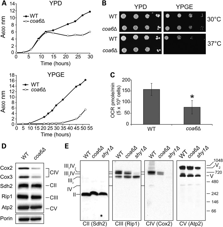
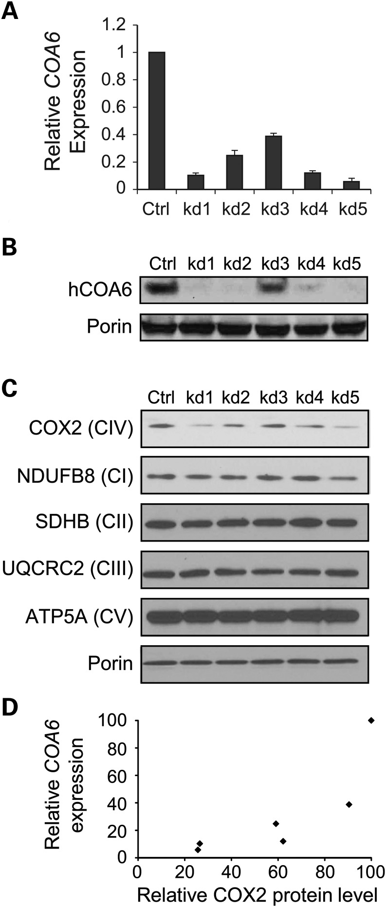
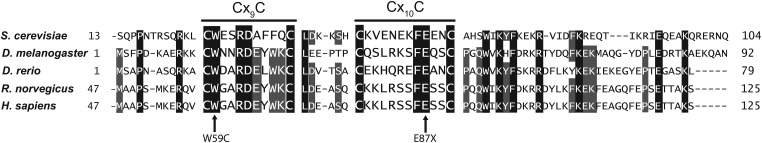
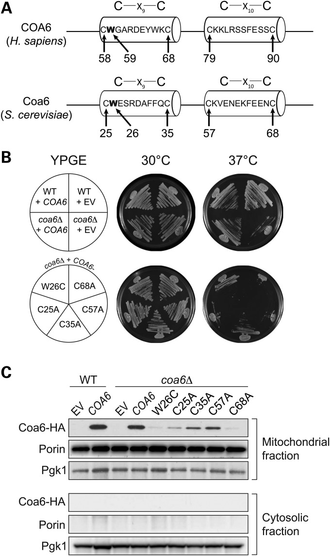
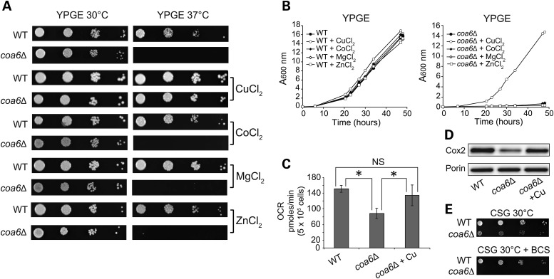
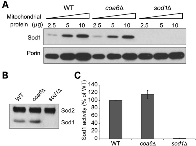
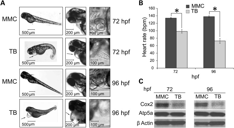
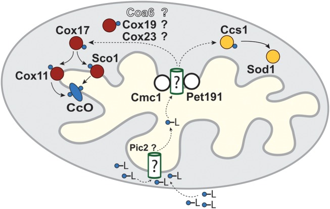
Mitochondrial respiratory chain biogenesis is orchestrated by hundreds of assembly factors, many of which are yet to be discovered. Using an integrative approach based on clues from evolutionary history, protein localization and human genetics, we have identified a conserved mitochondrial protein, C1orf31/COA6, and shown its requirement for respiratory complex IV biogenesis in yeast, zebrafish and human cells. A recent next-generation sequencing study reported potential pathogenic mutations within the evolutionarily conserved Cx₉CxnCx₁₀C motif of COA6, implicating it in mitochondrial disease biology. Using yeast coa6Δ cells, we show that conserved residues in the motif, including the residue mutated in a patient with mitochondrial disease, are essential for COA6 function, thus confirming the pathogenicity of the patient mutation. Furthermore, we show that zebrafish embryos with zfcoa6 knockdown display reduced heart rate and cardiac developmental defects, recapitulating the observed pathology in the human mitochondrial disease patient who died of neonatal hypertrophic cardiomyopathy. The specific requirement of Coa6 for respiratory complex IV biogenesis, its intramitochondrial localization and the presence of the Cx₉CxnCx₁₀C motif suggested a role in mitochondrial copper metabolism. In support of this, we show that exogenous copper supplementation completely rescues respiratory and complex IV assembly defects in yeast coa6Δ cells. Taken together, our results establish an evolutionarily conserved role of Coa6 in complex IV assembly and support a causal role of the COA6 mutation in the human mitochondrial disease patient.


Figures

Figure 1.
Figure 1.
Yeast coa6Δ cells exhibit reduced respiration and diminished CcO assembly. (A) WT and coa6Δ cells were precultured in YPD and inoculated in fresh YPD or YPGE liquid media at 30°C with a starting A600 of 0.1. Absorbance was measured at the indicated times at 600 nm. Data are representative of at least three independent measurements. (B) Serial dilutions of WT and coa6Δ cells were spotted on YPD and YPGE plates at 30°C and 37°C. Pictures were taken after 2–5 days of spotting. Data are representative of three independent experiments. (C) WT and coa6Δ cells were grown overnight in YPD at 30°C. After 18 h of growth, cells were harvested, washed, counted and resuspended in ethanol-containing respiratory medium. Basal oxygen consumption rate (OCR) was measured on half a million cells using an extracellular flux analyzer. Error bars represent mean ± SD (n = 10), * denotes statistically significant differences, P < 0.001, t-test. (D) Mitochondria were isolated from WT and coa6Δ cells grown to early stationary phase in YPD medium. Mitochondrial protein was extracted and analyzed by SDSPAGE/western blot. Subunit-specific antibodies were used to detect MRC complexes II–V. Porin was used as loading control. (E) Mitochondria from (D) and shy1Δ were solubilized in 1% digitonin, followed by BNPAGE/western blot of native MRC complexes. The shy1Δ cells lack CcO and thus were used as a control for CcO assembly. The stoichiometry and molecular weights of the supercomplexes are indicated.
Figure 2.
Figure 2.
Coa6 is required for the maintenance of MRC complex IV subunit (COX2) levels in human cells. (A) COA6 mRNA was quantified by qPCR on RNA extracted from MCH58 human fibroblasts infected either with an empty vector, pLKO.1 (Ctrl), or with one of the five independent shRNAs targeting COA6 (kd1–kd5). Values are reported as percent change in expression over control (Ctrl). Three replicates were used per hairpin (error bars represent S.D.). β-actin was used as an endogenous control in the qPCR. (B) Western blot detection of Coa6 protein abundance in Coa6 knockdown cell lines. (C) Western blot of individual subunits of MRC complexes (I–V) in mitochondria isolated from each of the COA6 knockdown cell lines. Western blot image is representative of three independent experiments. (D) Correlation between COA6 mRNA levels (from A) and COX2 levels (from C). COX2 levels were quantified by densitometry.
Figure 3.
Figure 3.
COA6 contains an evolutionarily conserved Cx9CxnCx10C motif. Sequence alignment of the conserved region of the human COA6 protein with its orthologs in yeast (Saccharomyces cerevisiae), fly (Drosophila melanogaster), fish (Danio rerio) and rat (Rattus norvegicus). The sequence alignment was performed using ClustalW. Horizontal lines above Cx9C and Cx10C residues show the conserved motif. Arrows indicate amino acid residues (tryptophan 59 and glutamic acid 87) that were mutated in a mitochondrial disease patient (6).
Figure 4.
Figure 4.
Conserved residues in the Cx9CxnCx10C motif are essential for its function. (A) Schematic representation of the Cx9CxnCx10C motif of human COA6 and yeast Coa6 proteins showing the location of cysteine and tryptophan residues (arrows) targeted for site-directed mutagenesis. (B) WT and coa6Δ cells transformed with empty vector (EV), COA6, or one of its mutant forms were streaked onto YPGE plates and incubated at 30°C or 37°C for 5 days before imaging. (C) Western blot analysis of mitochondrial and cytosolic extract from coa6Δ cells transformed with EV, HA-tagged COA6, and its mutant forms. Pgk1 is used as a cytosolic marker, and Porin is a mitochondrial marker.
Figure 5.
Figure 5.
Cu supplementation rescues respiratory and CcO assembly defects of coa6Δ. (A) Serially diluted WT and coa6Δ cells were spotted on YPGE plates in the presence and absence of 5 µm Cu, Co, Mg and Zn bivalent salts at 30°C and 37°C and allowed to grow for 4–5 days before imaging. (B) Growth of WT and coa6Δ cells in YPGE liquid media in the presence of 5 µm Cu, Co, Mg and Zn bivalent salts at 30°C. (C) Oxygen consumption rate (OCR) of WT, coa6Δ and coa6Δ supplemented with 5 µM CuSO4. Error bars represent mean ± SD (n = 6), *P < 0.05, t-test. NS, not significant. (D) Western blot of Cox2 in mitochondria isolated from WT, coa6Δ, and coa6Δ cells supplemented with 5 µm CuSO4. Porin is used as a loading control. (E) Serial dilutions of WT and coa6Δ strains grown in synthetic medium with a non-fermentable carbon source (2% glycerol; CSG) with or without 20 μm bathocuproinedisulfonic acid (BCS). The pictures were taken 5 days after seeding.
Figure 6.
Figure 6.
Mitochondrial Sod1 levels are unaltered in coa6Δ cells. (A) Mitochondria were isolated from the WT, coa6Δ and sod1Δ cells grown at 30°C in YPD to early stationary phase. Mitochondrial protein extract (2.5, 5 and 10 µg) was separated on 4–12% polyacrylamide gel and probed for Sod1 and porin by immunoblotting. Porin was used as a loading control. The blot is a representative of two independent experiments. (B) Sod activity in isolated mitochondria from the indicated strains was measured in an in-gel assay as described in Materials and Methods. Gel is representative of two independent experiments. (C) For quantification of the Sod activity in (B), the images were digitalized and densitometry performed using the ImageJ software.
Figure 7.
Figure 7.
Coa6 knockdown in zebrafish embryos results in cardiac defects. (A) Low- and high-resolution images of zebrafish embryos treated with mismatch control (MMC) and zfcoa6 translation blocking (TB) morpholinos at 72 and 96 h post fertilization (hpf). Arrows indicate pronounced pericardiac edema. High resolution images of the zebrafish heart in MMC and TB embryos were captured 72 and 96 hpf, where heart perimeter is outlined in MMC images to facilitate comparison to TB hearts. The images are representative of four independent experiments, where each experiment had between 150–250 embryos per morpholino. (B) Heart rate (beats/min) was measured in MMC and TB embryos at 72 and 96 hpf. Error bars represent mean ± SD and * indicates statistically significant differences, P < 0.001, t-test. (C) Western blot analysis of whole embryo protein extracts with indicated antibodies against CcO and complex V subunits. β actin is used as a loading control. The blot is representative of three independent experiments.
Figure 8.
Figure 8.
Schematic of the proposed mitochondrial copper transport pathways to CcO subunits. Cu (blue circles) enters the mitochondrial matrix bound to an unidentified ligand (-L) partially through the recently identified Pic2 protein. The Cu-L is stored in the matrix and exported to the intermembrane space to be delivered to CcO and Sod1 through a series of metallochaperones depicted in red and yellow, respectively. Only the final steps of Cu delivery mediated by Cox17, Cox11 and Sco1 are experimentally demonstrated. Dashed arrows indicate the hypothetical role of the several Cx9C containing proteins, including Coa6, in Cu transfer from a matrix Cu pool, across the inner mitochondrial membrane, to Cox17.

References

    1. Skladal D., Halliday J., Thorburn D.R. Minimum birth prevalence of mitochondrial respiratory chain disorders in children. Brain. 2003;126:1905–1912. - PubMed
    1. DiMauro S., Schon E.A. Mitochondrial respiratory-chain diseases. N. Engl. J. Med. 2003;348:2656–2668. - PubMed
    1. Fernández-Vizarra E., Tiranti V., Zeviani M. Assembly of the oxidative phosphorylation system in humans: what we have learned by studying its defects. Biochim. Biophys. Acta. 2009;1793:200–211. - PubMed
    1. Vafai S.B., Mootha V.K. Mitochondrial disorders as windows into an ancient organelle. Nature. 2012;491:374–383. - PubMed
    1. Chakravarti A., Clark A.G., Mootha V.K. Distilling pathophysiology from complex disease genetics. Cell. 2013;155:21–26. - PMC - PubMed

Publication types