

Resting brain activity varies with dream recall frequency between subjects
- PMID: 24549103
- PMCID: PMC4023156
- DOI: 10.1038/npp.2014.6
Resting brain activity varies with dream recall frequency between subjects
Abstract
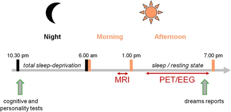
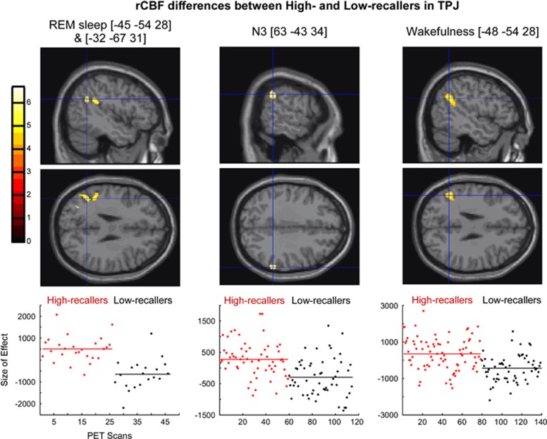
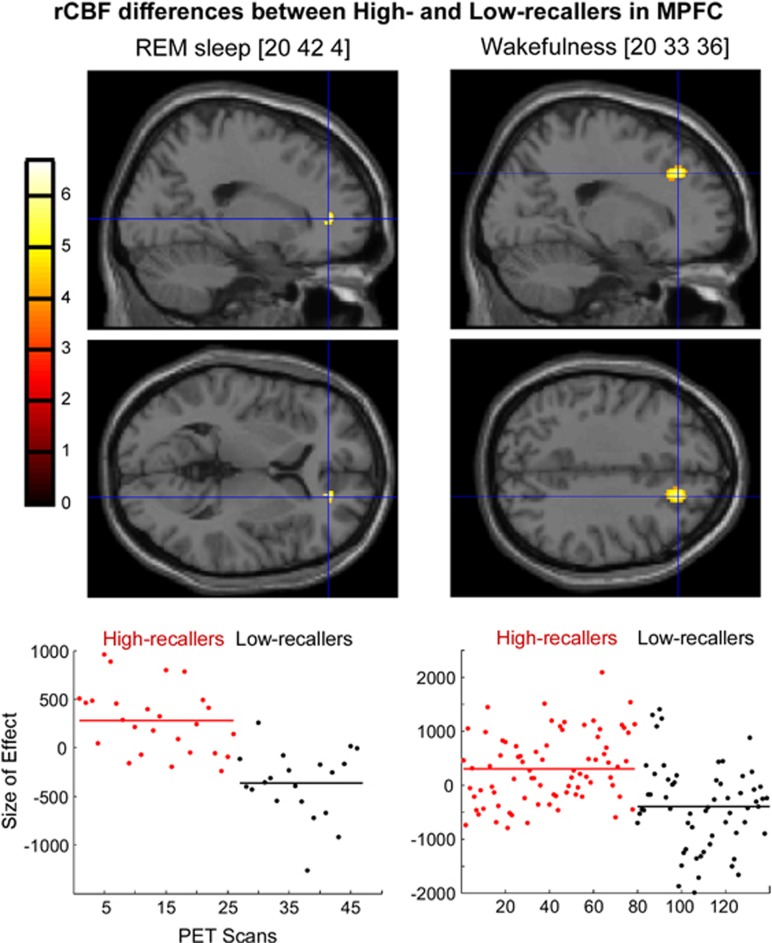
Dreaming is still poorly understood. Notably, its cerebral underpinning remains unclear. Neuropsychological studies have shown that lesions in the temporoparietal junction (TPJ) and/or the white matter of the medial prefrontal cortex (MPFC) lead to the global cessation of dream reports, suggesting that these regions of the default mode network have key roles in the dreaming process (forebrain 'dream-on' hypothesis). To test this hypothesis, we measured regional cerebral blood flow (rCBF) using [(15)O]H2O positron emission tomography in healthy subjects with high and low dream recall frequencies (DRFs) during wakefulness (rest) and sleep (rapid eye movement (REM) sleep, N2, and N3). Compared with Low recallers (0.5 ± 0.3 dream recall per week in average), High recallers (5.2 ± 1.4) showed higher rCBF in the TPJ during REM sleep, N3, and wakefulness, and in the MPFC during REM sleep and wakefulness. We demonstrate that the resting states of High recallers and Low recallers differ during sleep and wakefulness. It coheres with previous ERP results and confirms that a high/low DRF is associated with a specific functional organization of the brain. These results support the forebrain 'dream-on' hypothesis and suggest that TPJ and MPFC are not only involved in dream recall during wakefulness but also have a role in dreaming during sleep (production and/or encoding). Increased activity in the TPJ and MPFC might promote the mental imagery and/or memory encoding of dreams. Notably, increased activity in TPJ might facilitate attention orienting toward external stimuli and promote intrasleep wakefulness, facilitating the encoding of the dreams in memory.
Figures
References
-
- Aserinsky E, Kleitman N. Regularly occurring periods of eye motility, and concomitant phenomena, during sleep. Science. 1953;118:273–274. - PubMed
-
- Baars BJ, Ramsoy TZ, Laureys S. Brain, conscious experience and the observing self. Trends Neurosci. 2003;26:671–675. - PubMed
-
- Benoit O, Foret J. Le sommeil humain: bases expérimentales physiologiques et physiopathologiques. Masson: Paris; 1992. p. 197pp.
-
- Bischof M, Bassetti CL. Total dream loss: a distinct neuropsychological dysfunction after bilateral PCA stroke. Ann Neurol. 2004;56:583–586. - PubMed
-
- Blagrove M, Pace-Schott EF. Trait and neurobiological correlates of individual differences in dream recall and dream content. Int Rev Neurobiol. 2010;92:155–180. - PubMed
Publication types
MeSH terms
Substances
LinkOut - more resources
Full Text Sources
Other Literature Sources
Molecular Biology Databases
