Review
. 2014 Jun:26:132-41.
doi: 10.1016/j.conb.2014.01.015. Epub 2014 Feb 16.

Lineage origins of GABAergic versus glutamatergic neurons in the neocortex

Affiliations
Review

Lineage origins of GABAergic versus glutamatergic neurons in the neocortex

Oscar Marín et al. Curr Opin Neurobiol. 2014 Jun.

Abstract

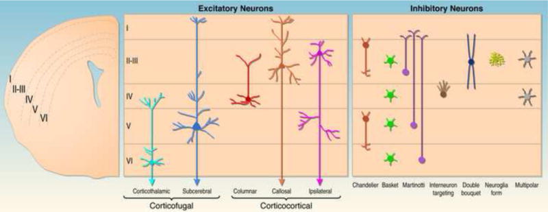
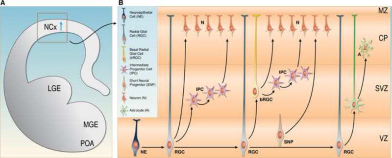
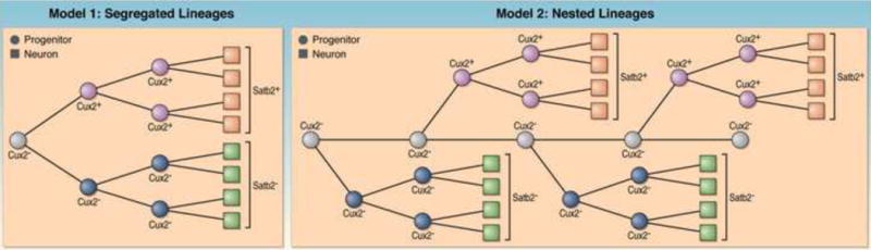
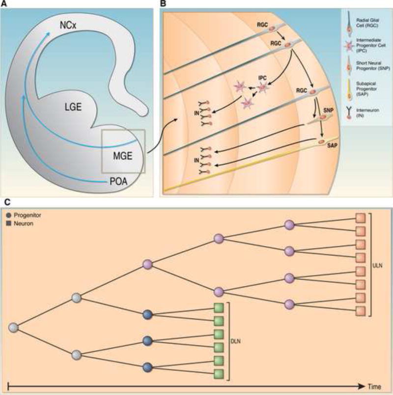
Neocortical circuits are assembled from subtypes of glutamatergic excitatory and GABAergic inhibitory neurons with divergent anatomical and molecular signatures and unique physiological properties. Excitatory neurons derive from progenitors in the pallium, whereas inhibitory neurons originate from progenitors in the subpallium. Both classes of neurons subsequently migrate along well-defined routes to their final target area, where they integrate into common neuronal circuits. Recent findings show that neuronal diversity within the lineages of excitatory and inhibitory neurons is in part already established at the level of progenitor cells before migration. This poses challenges for our understanding of how radial units of interconnected excitatory and inhibitory neurons are assembled from progenitors that are spatially segregated and diverse in nature.


Figures

Fig. 1
Fig. 1. Subtypes of glutamatergic excitatory and GABAergic inhibitory and their laminar distribution within the neocortex
On the right a coronal hemisection of the brain is shown. Neocortical cell layers I–VI are indicated. The middle panel shows the position of major subtypes of excitatory neurons within neocortical cell layers and their projection pattern. Note that the diagram is a simplification outlining the major laminar distribution of neuronal subtypes. For example, the majority of callosal projection neurons is located in layers II and III, but a significant subset is also found in deeper layers. The left panel shows the position of several subtypes of inhibitory interneurons within neocortical cell layers.
Fig. 2
Fig. 2. Subtypes of progenitor cells in the developing neocortex
(A) Coronal hemisection of the developing brain. The neocortex (NCx), the lateral ganglionic eminence (LGE), the medial ganglionic eminence (MGE), and the preoptic area (POA) are indicated. The blue arrow shows the migration route of excitatory neurons from the ventricular zone into the developing cortical wall. (B) Enlargement of the region boxed in (A) indicating different progenitor types. Neuroepithelial cells (NE) are present early in development and give rise to radial glial cells (RGCs). RGCs self-renew and give rise to neurons (N). RGCs also generate intermediate progenitors (IPCs) and short neural progenitors (SNPs), which divide further to generate neurons. In addition, RGCs give rise to basal radial glial cells (bRGCs), which generate via IPs additional neurons. At the end of neurogenesis, RGCs and bRGCs transform into astrocyte progenitors, which then generate astrocytes (A).
Fig. 3
Fig. 3. Models for the generation of subtypes of excitatory projection neurons
Two models are depicted that can explain how distinct progenitors generate Satb2+ callosal projection neurons and Satb2 subcerebral projection neurons. These are two extreme models and other models can be envisioned. Model 1: Segregated Lineages. An early progenitor generates two progenitor subtypes. These progenitors self-renew to amplify the progenitor pool and establish two independent progenitor lineages. One of the progenitor lineages is specified to produce Satb2 neurons that mostly populate deep layers, while the second progenitor lineage is specified to produce Satb2+ neurons that largely populate layers II–III but also reside in deeper layers. Model 2: Nested Lineages. A multipotent progenitor persists for an extended period of time and generates two progenitor subtypes. These progenitor subtypes proliferate to expand the two independent progenitor lineages. One of these progenitor subtypes generates Satb2+ neurons and the second one produces Satb2 neurons.
Fig. 4
Fig. 4. Generation of subtypes of interneurons
(A) Coronal hemisection of the developing brain. The blue arrow indicates the migration route of interneurons from the medial ganglionic eminence (MGE) to the neocortex (NCx). The lateral ganglionic eminence (LGE) is also depicted. (B) Enlargement of the region boxed in (A) indicating different progenitor types. Radial glial cells (RGCs) that were derived from neuroepithelial cells self renew and generate intermediate progenitors (IPCs), short neural progenitors (SNPs) and subapical progenitors (SAPs), all of which further proliferate to generate interneurons (INs). (C) Model for the generation of interneurons that populate deep layers (DLN) and upper layers (ULN) of the neocortex. In this model, distinct progenitors exist for DLN and ULN but their relative abundance changes over developmental time. Note that most of the data regarding the diversity of the progenitor pool for interneurons were obtained with studies on the LGE, which is not depicted here.

References

    1. Greig LC, Woodworth MB, Galazo MJ, Padmanabhan H, Macklis JD. Molecular logic of neocortical projection neuron specification, development and diversity. Nat Rev Neurosci. 2013;14:755–769. - PMC - PubMed
    1. Franco SJ, Muller U. Shaping our minds: stem and progenitor cell diversity in the mammalian neocortex. Neuron. 2013;77:19–34. - PMC - PubMed
    1. DeFelipe J, Lopez-Cruz PL, Benavides-Piccione R, Bielza C, Larranaga P, Anderson S, Burkhalter A, Cauli B, Fairén A, Feldmeyer D, et al. New insights into the classification and nomenclature of cortical GABAergic interneurons. Nat Rev Neurosci. 2013;14:202–216. - PMC - PubMed
    1. Rakic P. Specification of cerebral cortical areas. Science. 1988;241:170–176. - PubMed
    1. Rakic P. The radial edifice of cortical architecture: from neuronal silhouettes to genetic engineering. Brain Res Rev. 2007;55:204–219. - PMC - PubMed

Publication types