. 2014 May;23(5):618-26.
doi: 10.1002/pro.2444. Epub 2014 Mar 11.

Structural insight into the dimerization of human protein disulfide isomerase

Affiliations

Structural insight into the dimerization of human protein disulfide isomerase

Sara Bastos-Aristizabal et al. Protein Sci. 2014 May.

Abstract

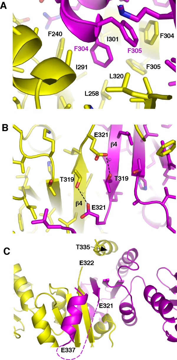
Protein disulfide isomerases (PDIs) are responsible for catalyzing the proper oxidation and isomerization of disulfide bonds of newly synthesized proteins in the endoplasmic reticulum (ER). Here, it is shown that human PDI (PDIA1) dimerizes in vivo and proposed that the dimerization of PDI has physiological relevance by autoregulating its activity. The crystal structure of the dimeric form of noncatalytic bb' domains of human PDIA1 determined to 2.3 Å resolution revealed that the formation of dimers occludes the substrate binding site and may function as a mechanism to regulate PDI activity in the ER.

Keywords: crystal structure; dimerization; endoplasmic reticulum; protein disulfide isomerase; thioredoxin-like domain.


Figures

Figure 1
Figure 1
In vivo dimerization of PDIA1. co-IPs with anti-myc (lanes 1–3) and with anti-FLAG (lane 4–6) were analyzed by SDS-PAGE followed by Western blot using anti-myc (A) and anti-FLAG (B). Asterisks indicate bands that belong to the heavy chains of the antibodies used in the immunoprecipitation.
Figure 2
Figure 2
PDIA1 dimerization occurs in the cells. co-IPs with anti-myc (lanes 1, 2) and with anti-FLAG (lane 3, 4) were analyzed by SDS-PAGE and Western blot using anti-myc (A) and anti-FLAG (B). Indicated with an asterisk are the bands that belong to the antibodies used in the immunoprecipitation.
Figure 3
Figure 3
PDIA1 dimerization is disulfide independent. co-IPs with anti-myc (lanes 1–4) and with anti-FLAG (lane 5–8) were analyzed by SDS-PAGE followed by Western blot using anti-myc (A) and anti-FLAG (B). Cross-reactive bands from antibody proteins are marked by an asterisk.
Figure 4
Figure 4
Crystal structure of PDIA1 bb′ dimer. A ribbon diagram showing PDIA1 dimer in two different orientations. The dimer is colored by chain in which one is represented by yellow and the other one in magenta and the domains are labeled.
Figure 5
Figure 5
Comparison of the b′ substrate-binding sites in dimeric and monomeric structures. (A) F304 and F305 of the b′ domain occupy the substrate-binding site of another b′ domain in the dimeric structure. (B) L343 and W347 of the X-linker interact with monomeric b′ domain (PDB code 2BJX).
Figure 6
Figure 6
Dimerization does not affect relative orientation of the b and b′ domain. Orientation of the bb′ domains in the dimeric form (in yellow) is very similar to that in reduced monomeric PDIA1 (PDB code 4EKZ; in green) (A) and differs in oxidized PDIA1 (PDB code 4EL1; in blue) (B).
Figure 7
Figure 7
Structural determinants of PDIA1 bb′ dimerization. Dimer forms via interactions of one molecule (yellow) with another (magenta) through their hydrophobic pockets. Dimerization is mediated by hydrophobic interactions (A), polar interactions (B) and swapping of the C-terminal α-helices (C).

References

    1. Appenzeller-Herzog C, Ellgaard L. The human PDI family: versatility packed into a single fold. Biochim Biophys Acta. 2008;1783:535–548. - PubMed
    1. Ellgaard L, Ruddock LW. The human protein disulphide isomerase family: substrate interactions and functional properties. EMBO Rep. 2005;6:28–32. - PMC - PubMed
    1. Karala AR, Lappi AK, Saaranen MJ, Ruddock LW. Efficient peroxide-mediated oxidative refolding of a protein at physiological pH and implications for oxidative folding in the endoplasmic reticulum. Antiox Redox Signal. 2009;11:963–970. - PubMed
    1. Benham AM. The protein disulfide isomerase family: key players in health and disease. Antiox Redox Signal. 2012;16:781–789. - PubMed
    1. Tian G, Xiang S, Noiva R, Lennarz WJ, Schindelin H. The crystal structure of yeast protein disulfide isomerase suggests cooperativity between its active sites. Cell. 2006;124:61–73. - PubMed

Publication types

MeSH terms

LinkOut - more resources