. 2014 Feb 18;5(1):e00931-13.
doi: 10.1128/mBio.00931-13.

Reovirus forms neo-organelles for progeny particle assembly within reorganized cell membranes

Reovirus forms neo-organelles for progeny particle assembly within reorganized cell membranes

Isabel Fernández de Castro et al. mBio. .

Erratum in

Abstract

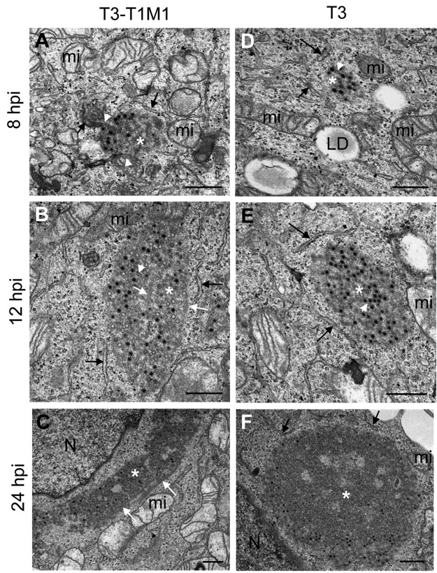
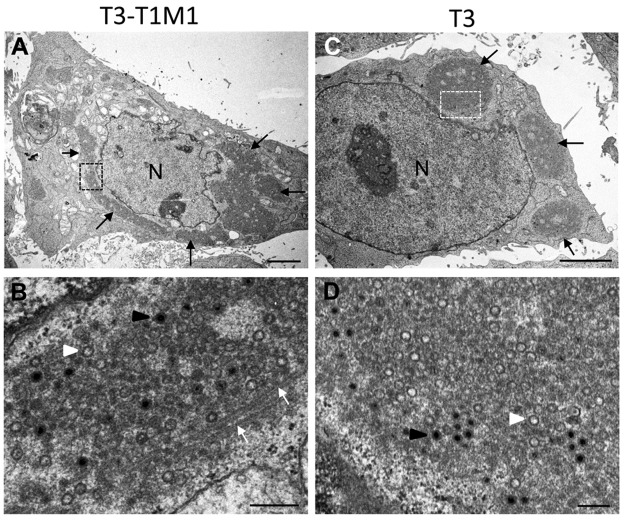
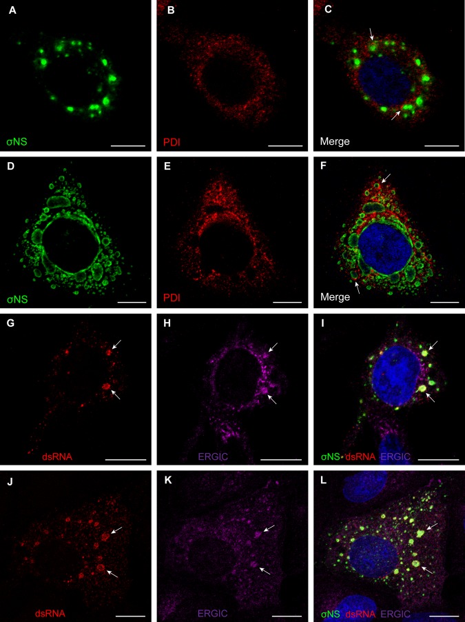
Most viruses that replicate in the cytoplasm of host cells form neo-organelles that serve as sites of viral genome replication and particle assembly. These highly specialized structures concentrate viral replication proteins and nucleic acids, prevent the activation of cell-intrinsic defenses, and coordinate the release of progeny particles. Despite the importance of inclusion complexes in viral replication, there are key gaps in the knowledge of how these organelles form and mediate their functions. Reoviruses are nonenveloped, double-stranded RNA (dsRNA) viruses that serve as tractable experimental models for studies of dsRNA virus replication and pathogenesis. Following reovirus entry into cells, replication occurs in large cytoplasmic structures termed inclusions that fill with progeny virions. Reovirus inclusions are nucleated by viral nonstructural proteins, which in turn recruit viral structural proteins for genome replication and particle assembly. Components of reovirus inclusions are poorly understood, but these structures are generally thought to be devoid of membranes. We used transmission electron microscopy and three-dimensional image reconstructions to visualize reovirus inclusions in infected cells. These studies revealed that reovirus inclusions form within a membranous network. Viral inclusions contain filled and empty viral particles and microtubules and appose mitochondria and rough endoplasmic reticulum (RER). Immunofluorescence confocal microscopy analysis demonstrated that markers of the ER and ER-Golgi intermediate compartment (ERGIC) codistribute with inclusions during infection, as does dsRNA. dsRNA colocalizes with the viral protein σNS and an ERGIC marker inside inclusions. These findings suggest that cell membranes within reovirus inclusions form a scaffold to coordinate viral replication and assembly.

Importance: Viruses alter the architecture of host cells to form an intracellular environment conducive to viral replication. This step in viral infection requires the concerted action of viral and host components and is potentially vulnerable to pharmacological intervention. Reoviruses form large cytoplasmic replication sites called inclusions, which have been described as membrane-free structures. Despite the importance of inclusions in the reovirus replication cycle, little is known about their formation and composition. We used light and electron microscopy to demonstrate that reovirus inclusions are membrane-containing structures and that the endoplasmic reticulum (ER) and the ER-Golgi intermediate compartment interact closely with these viral organelles. These findings enhance our understanding of the cellular machinery usurped by viruses to form inclusion organelles and complete an infectious cycle. This information, in turn, may foster the development of antiviral drugs that impede this essential viral replication step.


Figures

FIG 1
FIG 1
Ultrastructure of reovirus inclusions in HeLa cells. HeLa cells were infected with reovirus strain T3-T1M1 (A to C) or T3 (D to F) and fixed at 8, 12, or 24 hpi. Ultrathin (~60- to 70-nm) sections were imaged by TEM. Inclusions are marked with white asterisks, RER cisternae are marked with black arrows, smooth membranes inside inclusions are marked with white arrowheads, and coated microtubules are marked with white arrows. T3-T1M1 inclusions at 8 (A), 12 (B), and 24 (C) hpi and T3 inclusion at 8 (D), 12 (E), and 24 (F) hpi are shown. LD, lipid droplet; mi, mitochondria; N, nucleus. Scale bars: 0.5 µm.
FIG 2
FIG 2
TEM of HeLa cells infected with reovirus strain T3-T1M1 or T3 at 24 hpi. Low (A) and high (B) magnifications of cells infected with T3-T1M1 are shown. (A) Perinuclear inclusion (black arrows). (B) Enlargement of highlighted area in panel A. The inclusion contains filled viral particles (black arrowhead), empty viral particles (white arrowhead), and coated microtubules (white arrows). (C, D) Low (C) and high (D) magnifications of cells infected with T3. In panel C, perinuclear inclusions are marked with black arrows. Panel D is an enlargement of the boxed area in panel C. The inclusion contains filled (black arrowhead) and empty (white arrowhead) particles. Scale bars: 3 µm in panels A and C, 0.25 µm in panels B and D.
FIG 3
FIG 3
TEM of viral inclusions in T3-T1M1-infected HeLa cells. Inclusions are marked with white asterisks. (A) Viral particles (arrows) in small inclusions attached to membranes in a region containing RER and the Golgi compartment at 8 hpi. (B) Smooth membranes (white arrowheads) inside an inclusion surrounded by RER at 8 hpi. (C) Inclusion at 12 hpi surrounded by mitochondria and ER membranes. Some RER elements close to the inclusion (arrow on the right) appear to be connected to a sheet of cubic membranes (black asterisk). (D) Inclusion at 24 hpi filled with coated microtubules (white arrows). (E) Higher magnification of a different inclusion with viral particles attached to coated (~60 nm, white arrows) but not uncoated (~30 nm, black arrows) microtubules. N, nucleus; C, centriole. Scale bars: 0.25 µm.
FIG 4
FIG 4
Reovirus inclusions codistribute with ER and ERGIC elements. (A to F) HeLa cells were infected with T3-T1M1 for 12 h (A to C) or 24 h (D to F). Cells were fixed; permeabilized; stained for σNS (green), PDI (red), or nuclei (blue); and visualized by confocal microscopy. Arrows indicate viral inclusions associated with RER elements on the periphery. (G to L) HeLa cells (G to I) and MDCK cells (J to L) were infected with T3-T1M1 for 12 h; fixed; permeabilized; stained for dsRNA (red), ERGIC-53 (magenta), σNS (green), or nuclei (blue); and visualized by confocal microscopy. Arrows indicate viral inclusions that contain dsRNA, the ERGIC, and σNS. Scale bars: 10 µm.
FIG 5
FIG 5
3D model of reovirus inclusions in HeLa cells. HeLa cells were infected with T3-T1M1 and fixed at 12 hpi. The inclusion was visualized by TEM of serial sections, 3D reconstruction, and image processing. (A) Mitochondria (red) surround a network of membranes (yellow). RER (brown) and nuclear envelope (blue) are adjacent to the inclusion. Filled viral particles (black), empty viral particles (white), and microtubules (green) are integrated into the inclusion membrane network. (B) Rotation of the same reconstruction showing contacts between the inclusion membranes and mitochondria (arrows). The volume has been superimposed onto the 2D image of one of the sections in the series.
FIG 6
FIG 6
3D model of reovirus inclusions in MDCK cells. MDCK cells were infected with T3-T1M1 (A and C to E) or T3 (B) and fixed at 24 hpi. Membranes (yellow), mitochondria (red), RER (light brown), microtubules (green), filled viral particles (black), and empty viral particles (white) are shown. (A) Image reconstruction of an inclusion formed in T3-T1M1-infected cells. RER membranes surround and incorporate into the inclusion membrane network that contains numerous mature virions, fewer empty particles, and microtubules. (B) Image reconstruction of an inclusion formed in T3-infected cells. Most of the viral particles inside the membranous web are empty. No microtubules were observed inside or around the inclusion. Mitochondria, RER, and lipid droplets (large white structures) surround the inclusion, which is in contact with the nuclear envelope (blue). (C) Image reconstruction of an inclusion close to the plasma membrane (brown) in a cell infected with T3-T1M1. The plasma membrane from another cell is colored blue. The inclusion contains membranes (yellow) and filled (black) and empty (white) viral particles. Peripheral RER elements, microtubules, and viral particles are in contact with the cytosolic face of the plasma membrane. Enlargements of this region are shown in panels D and E. Viral particles attached to RER, microtubules, and the plasma membrane (arrows) are visible.

References

    1. Miller S, Krijnse-Locker J. 2008. Modification of intracellular membrane structures for virus replication. Nat. Rev. Microbiol. 6:363–374. 10.1038/nrmicro1890 - DOI - PMC - PubMed
    1. Paul D, Bartenschlager R. 2013. Architecture and biogenesis of plus-strand RNA virus replication factories. World J. Virol. 2:32–48. 10.5501/wjv.v2.i2.32 - DOI - PMC - PubMed
    1. Paul D, Hoppe S, Saher G, Krijnse-Locker J, Bartenschlager R. 2013. Morphological and biochemical characterization of the membranous hepatitis C virus replication compartment. J. Virol. 87:10612–10627. 10.1128/JVI.01370-13 - DOI - PMC - PubMed
    1. Novoa RR, Calderita G, Arranz R, Fontana J, Granzow H, Risco C. 2005. Virus factories: associations of cell organelles for viral replication and morphogenesis. Biol. Cell 97:147–172. 10.1042/BC20040058 - DOI - PMC - PubMed
    1. de Castro IF, Volonté L, Risco C. 2013. Virus factories: biogenesis and structural design. Cell. Microbiol. 15:24–34. 10.1111/cmi.12029 - DOI - PMC - PubMed

Publication types