. 2014 Mar;141(5):1085-94.
doi: 10.1242/dev.103333.

Developmental stratification of the mammary epithelium occurs through symmetry-breaking vertical divisions of apically positioned luminal cells

Affiliations

Developmental stratification of the mammary epithelium occurs through symmetry-breaking vertical divisions of apically positioned luminal cells

Robert J Huebner et al. Development. 2014 Mar.

Abstract

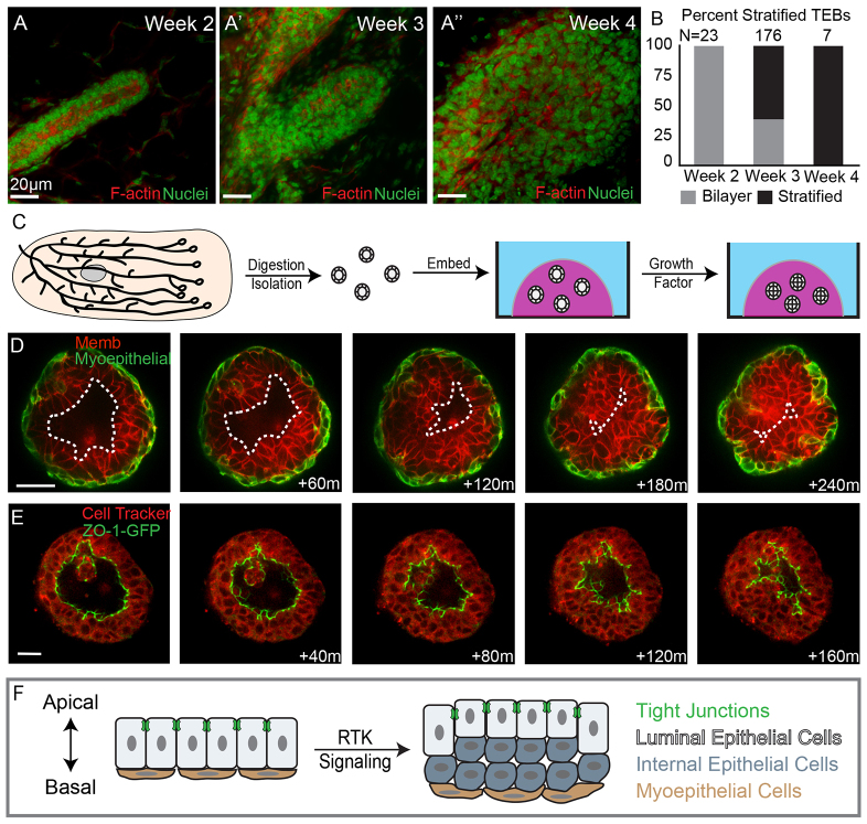
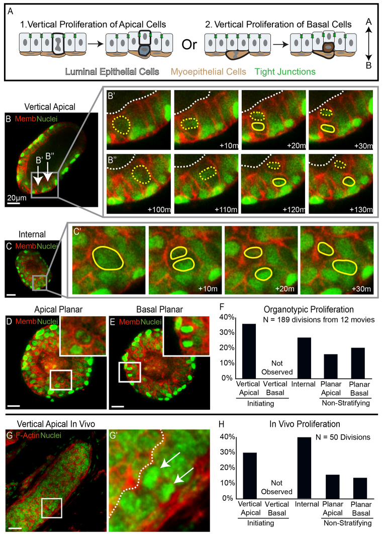
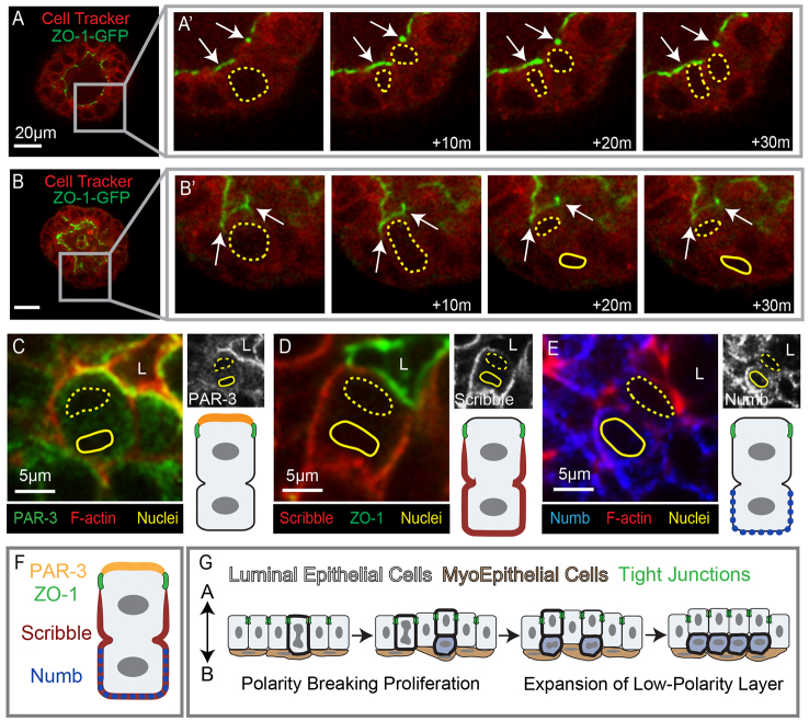
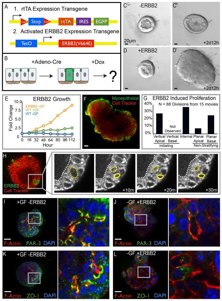
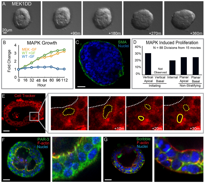
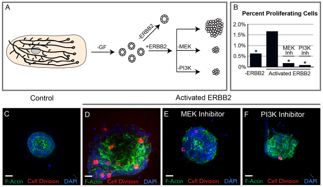
Mammary ducts are elongated during development by stratified epithelial structures, known as terminal end buds (TEBs). TEBs exhibit reduced apicobasal polarity and extensive proliferation. A major unanswered question concerns the mechanism by which the simple ductal epithelium stratifies during TEB formation. We sought to elucidate this mechanism using real-time imaging of growth factor-induced stratification in 3D cultures of mouse primary epithelial organoids. We hypothesized that stratification could result from vertical divisions in either the apically positioned luminal epithelial cells or the basally positioned myoepithelial cells. Stratification initiated exclusively from vertical apical cell divisions, both in 3D culture and in vivo. During vertical apical divisions, only the mother cell retained tight junctions and segregated apical membranes. Vertical daughter cells initiated an unpolarized cell population located between the luminal and myoepithelial cells, similar to the unpolarized body cells in the TEB. As stratification and loss of apicobasal polarity are early hallmarks of cancer, we next determined the cellular mechanism of oncogenic stratification. Expression of activated ERBB2 induced neoplastic stratification through analogous vertical divisions of apically positioned luminal epithelial cells. However, ERBB2-induced stratification was accompanied by tissue overgrowth and acute loss of both tight junctions and apical polarity. Expression of phosphomimetic MEK (MEK1DD), a major ERBB2 effector, also induced stratification through vertical apical cell divisions. However, MEK1DD-expressing organoids exhibited normal levels of growth and retained apicobasal polarity. We conclude that both normal and neoplastic stratification are accomplished through receptor tyrosine kinase signaling dependent vertical cell divisions within the luminal epithelial cell layer.

Keywords: Apicobasal polarity; Breast cancer; Epithelial development; Mammary gland; Mouse.


Figures

Fig. 1.
Fig. 1.
Mammary stratification generates an internal population of luminal epithelial cells lacking tight junctions. (A-A′) Terminal end buds from 2- to 4-week-old mice were stained for F-actin (red) and nuclei (green). (B) Quantification of bilayered (gray) and stratified (black) end buds at 2, 3 and 4 weeks postnatal. (C) A schematic depicting organotypic culture. (D) Still images from a movie of an organoid undergoing stratification. Luminal epithelial cells are labeled red with a membrane localized tdTomato and myoepithelial cells are labeled green with a genetically encoded green fluorescent protein (GFP). The dashed line highlights the boundary of the luminal space. (E) Still images of an organoid undergoing stratification with ZO-1-GFP marking tight junctions and Cell Tracker Red staining the cytosol. (F) A cartoon depiction of mammary epithelial stratification showing the generation of an internal luminal epithelial cell population lacking tight junctions. Scale bars: 20 μm.
Fig. 2.
Fig. 2.
Vertical apical cell divisions initiated mammary stratification. (A) A schematic depicting the alternatives of stratification initiation by vertical apical versus vertical basal proliferation. (B-E) Frames from movies of organoids expressing nuclear (green) and membrane (red) markers were collected to visualize proliferation in real time. (B) An image of an organoid before stratification. The arrows highlight apical cells that undergo vertical proliferation in B′ and B′. (B′,B′) Movie frames showing two vertical apical cell divisions, each producing one apical mother cell and one internal daughter cell. Yellow dashed lines show apical mother cells, solid yellow lines highlight internal daughter cells and dashed white lines represent the border of the lumen. (C) An organoid that is partially stratified with inset on an internal cell that divides in C′. (C′) Frames showing division of the internal cell highlighted in C; solid yellow lines outline the dividing nuclei. (D,E) An image of planar proliferation of apical and basal cells, respectively, insets on dividing cells. (F) Quantification of the types of proliferation observed in organotypic culture. (G) A terminal end bud from 3-week-old mouse stained for F-actin (red) and nuclei (green). The inset highlights a vertical apical division. (G′) An enlarged image of the inset from G; the arrows point to dividing nuclei and the dashed white line shows the border of the lumen. (H) Quantification of the types of proliferation observed in vitro. Scale bars: 20 μm.
Fig. 3.
Fig. 3.
Vertical apical cell divisions result in polarity loss for internal daughter cells. (A-B′) Frames from movies of a ZO-1-GFP-expressing organoid (green) counterstained with Cell Tracker Red. (A) An organoid undergoing stratification; the inset highlights a cell that undergoes planar apical proliferation in A′. (A′) Magnification of the inset from A; arrows point to tight junctions, which were apically localized during apical proliferation. Yellow dashed lines highlight both the mother and the daughter cell. (B) A still image of an organoid with inset on an apical cell before the vertical division shown in B′. (B′) Images show the asymmetric localization of ZO-1 to the apically positioned mother cell during vertical apical cell division. Yellow dashed lines emphasize the mother cell; solid lines show the internal daughter cell and arrows point to the tight junctions. (C-E) Immunofluorescence staining of apical cells during vertical divisions. The luminal space is marked L, apical nuclei are highlighted with dashed yellow lines and internal nuclei are shown with solid yellow lines. Black and white images of the respective polarity proteins are shown in the upper right and a schematic depiction of the protein localization is included in the bottom right. (C) PAR-3 was asymmetrically localized to the apical membrane of the mother cell during vertical division. (D) The basolateral polarity protein scribble was excluded from the ZO-1 defined apical domain but was present on all other basolateral surfaces. (E) Numb was enriched in the basolateral membranes of the daughter cell. (F) A cartoon depiction of the asymmetric localization of polarity proteins during vertical apical cell divisions. (G) A schematic depicting the cellular mechanism of mammary stratification initiating with polarity breaking vertical apical cell divisions followed by expansion of the low-polarity internal cell layer. Scale bars: 20 μm unless otherwise noted.
Fig. 4.
Fig. 4.
Oncogenic stratification shares a conserved cellular mechanism with developmental stratification. (A) Schematic of the transgenes used for conditional and mosaic expression of activated ERBB2. (B) Cartoon depicting the protocol followed to drive ERBB2 expression. (C-D′) Still images from time-lapse movies of unstimulated organoids in the presence or absence of ERBB2 expression. (C,C′) Organoids remained bilayered in the absence of ERBB2. (D,D′) Expression of ERBB2 in 80-90% of cells resulted in rapid stratification and growth. (E) Organoid growth rates were quantified as the increase in organoid area normalized to the initial organoid size. Growth curves for organoids expressing activated ERBB2 without exogenous growth factor are shown in orange, for control organoids with growth factor are shown in green, and for control organoids without exogenous growth factor are shown in blue. (F) Organoids expressing ERBB2 in 80-90% of cells were fixed and stained for smooth muscle actin (SMA, green), 3 days after gene activation. SMA, a marker of myoepithelial cells, showed that ERBB2-induced stratification increased the number of luminal epithelial cell layers. (G) Quantification of the types of cell divisions observed in ERBB2-expressing organoids. (H) Mosaic expression of ERBB2 in 1-10% of cells allowed observation of individual cellular responses to oncogene activation. ERBB2-expressing cells were labeled with GFP (green) and Cell Tracker Red was used to infer nuclear location. ERBB2-induced stratification initiated from vertical apical cell divisions. (I-L) Tissue was fixed and stained 3 days after addition of growth factor or activation of ERBB2 in 80-90% of cells. (I,J) Apical polarity was visualized by staining for PAR-3 (green) as well as F-actin (red) and nuclei (blue). (I) Polarized apical membranes were present at the apical surfaces of tissue treated with growth factor. (J) Tissue expressing ERBB2 lacked membrane localized PAR-3. (K,L) ZO-1 staining (green) was used to observe tight junctions; tissue was also stained for F-actin (red) and nuclei (blue). (K) Growth factor-treated organoids had ZO-1 localized to apical membranes. (L) Tight junctions were absent from apical membranes in cells expressing ERBB2; the inset shows a region of tissue lacking tight junctions. Scale bars: 20 μm.
Fig. 5.
Fig. 5.
MEK1DD-induced stratification through vertical apical cell divisions. (A) Still images from a time-lapse movie of organoids expressing a phosphomimetic form of MEK (MEK1DD). MEK1DD expression was sufficient to drive stratification. (B) Growth curves for organoids expressing MEK1DD without exogenous growth factor are shown in orange, for control organoids with growth factor in green and for organoids without exogenous growth factor in blue. (C) MEK1DD-expressing organoids were stained for SMA (green) and nuclei (blue) to visualize the myoepithelial cells. (D) Quantification of the types of proliferation observed in response to MEK1DD expression. (B) MEK1DD-induced stratification was initiated by vertical apical cell divisions; organoids were stained with Cell Tracker Red to infer nuclear position. (F) Organoids were stained for PAR-3 (green) F-actin (red) and nuclei (blue) to visualize apical polarity. (G) Organoids were stained for scribble (green) F-actin (red) and nuclei (blue) to visualize basolateral polarity. Scale bars: 20 μm.
Fig. 6.
Fig. 6.
MAPK and PI3 kinase are both required for ERBB2-induced proliferation. (A) Organoids were allocated to control and ERBB2-expressing experimental groups. A subset of the ERBB2-expressing organoids were treated with either MEK or PI3 kinase inhibitor. (B) Organoids were fixed 3 days after ERBB2 activation and stained for the proliferation marker PH3 (red), DAPI (blue) and F-actin (green). Quantification of the effect of MEK and PI3 kinase inhibitors on organoids expressing mutant ERBB2. Percent proliferation was quantified as the number of PH3-positive cells divided by the total number of cells per organoid. Stars highlight conditions that were significantly different by ANOVA (P<0.001) from the activated ERBB2 mutant. (B) Control organoids displayed a low level of proliferation. (C) ERBB2 activation increased the number of PH3-positive cells. (C) MEK inhibition with U0126 (U) decreased proliferation below the level observed in control organoids. (D) Inhibition of the AKT pathway with the PI3 kinase inhibitor LY294002 (LY) decreased proliferation below the level of control organoids. Scale bars: 20 μm.

Similar articles

Cited by

References

    1. Andrew D. J., Ewald A. J. (2010). Morphogenesis of epithelial tubes: Insights into tube formation, elongation, and elaboration. Dev. Biol. 341, 34–55 - PMC - PubMed
    1. Aranda V., Haire T., Nolan M. E., Calarco J. P., Rozenberg A. Z., Fawcett J. P., Pawson T., Muthuswamy S. K. (2006). Par6-aPKC uncouples ErbB2 induced disruption of polarized epithelial organization from proliferation control. Nat. Cell Biol. 8, 1235–1245 - PubMed
    1. Belteki G., Haigh J., Kabacs N., Haigh K., Sison K., Costantini F., Whitsett J., Quaggin S. E., Nagy A. (2005). Conditional and inducible transgene expression in mice through the combinatorial use of Cre-mediated recombination and tetracycline induction. Nucleic Acids Res. 33, e51 - PMC - PubMed
    1. Bryant D. M., Mostov K. E. (2008). From cells to organs: building polarized tissue. Nat. Rev. Mol. Cell Biol. 9, 887–901 - PMC - PubMed
    1. Edelstein A., Amodaj N., Hoover K., Vale R., Stuurman N. (2010). Computer control of microscopes using microManager. Curr. Protoc. Mol. Biol. 92, 14.20.1–14.20.17 - PMC - PubMed

Publication types