. 2014 Mar;141(5):1120-8.
doi: 10.1242/dev.101808.

Molecular identification of venous progenitors in the dorsal aorta reveals an aortic origin for the cardinal vein in mammals

Affiliations

Molecular identification of venous progenitors in the dorsal aorta reveals an aortic origin for the cardinal vein in mammals

Henrik Lindskog et al. Development. 2014 Mar.

Abstract

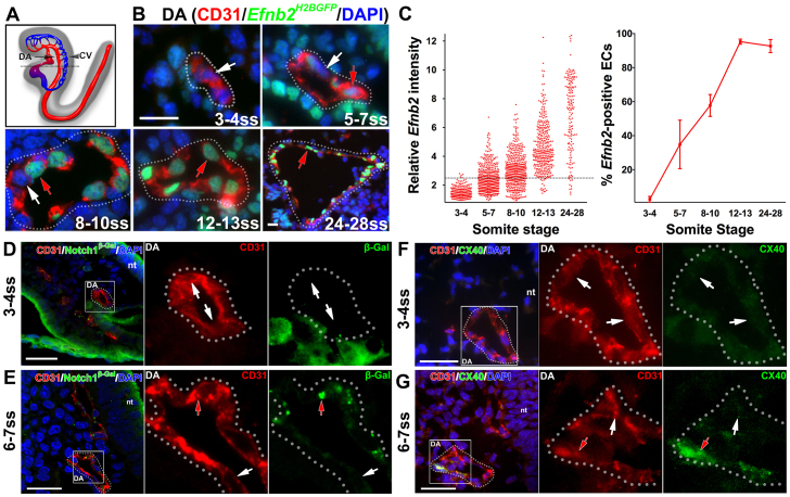
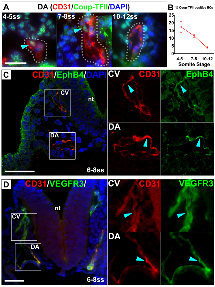
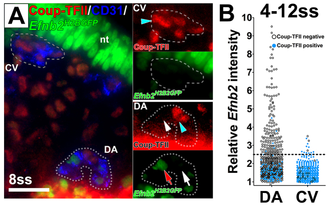
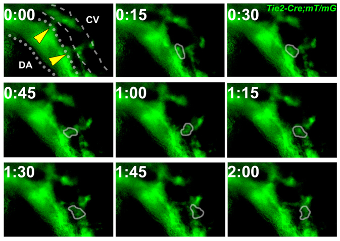
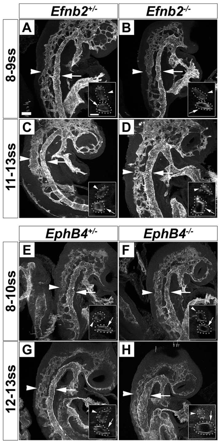
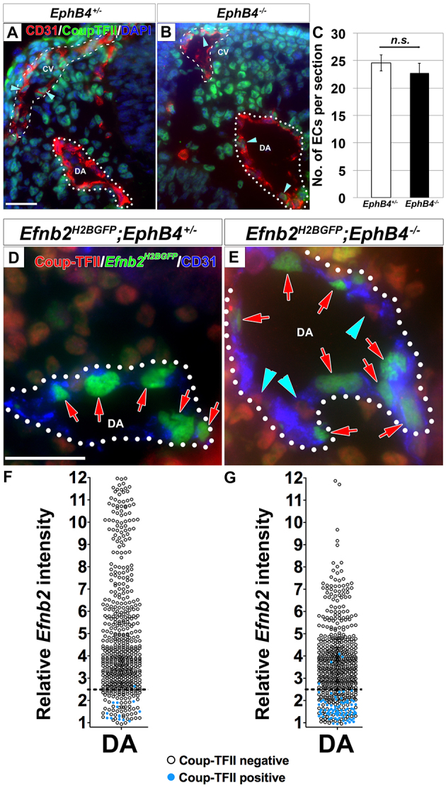
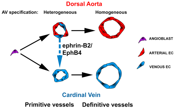
Coordinated arterial-venous differentiation is crucial for vascular development and function. The origin of the cardinal vein (CV) in mammals is unknown, while conflicting theories have been reported in chick and zebrafish. Here, we provide the first molecular characterization of endothelial cells (ECs) expressing venous molecular markers, or venous-fated ECs, within the emergent dorsal aorta (DA). These ECs, expressing the venous molecular markers Coup-TFII and EphB4, cohabited the early DA with ECs expressing the arterial molecular markers ephrin B2, Notch and connexin 40. These mixed ECs in the early DA expressed either the arterial or venous molecular marker, but rarely both. Subsequently, the DA exhibited uniform arterial markers. Real-time imaging of mouse embryos revealed EC movement from the DA to the CV during the stage when venous-fated ECs occupied the DA. We analyzed mutants for EphB4, which encodes a receptor tyrosine kinase for the ephrin B2 ligand, as we hypothesized that ephrin B2/EphB4 signaling may mediate the repulsion of venous-fated ECs from the DA to the CV. Using an EC quantification approach, we discovered that venous-fated ECs increased in the DA and decreased in the CV in the mutants, whereas the rest of the ECs in each vessel were unaffected. This result suggests that the venous-fated ECs were retained in the DA and missing in the CV in the EphB4 mutant, and thus that ephrin B2/EphB4 signaling normally functions to clear venous-fated ECs from the DA to the CV by cell repulsion. Therefore, our cellular and molecular evidence suggests that the DA harbors venous progenitors that move to participate in CV formation, and that ephrin B2/EphB4 signaling regulates this aortic contribution to the mammalian CV.

Keywords: Angiogenesis; Arterial-venous differentiation; Coup-TFII; EphB4; Ephrin B2; Mouse; Notch; Vascular development.


Figures

Fig. 1.
Fig. 1.
The dorsal aorta transiently contained both arterial marker-positive and -negative endothelial cells. (A-C) Arterial marker Efnb2 expression in cross-sections of the developing DA using the Efnb2H2BGFP/+ reporter. (A) Schematic lateral view of the DA (arrow) and the CV (arrowhead) at E8.5. The dotted black line indicates the level of cross-sections in B. (B) At 3-4 ss, the DA was composed of ECs without detectable Efnb2 expression. At 5-7 ss and 8-10 ss, both Efnb2-positive (red arrows) and -negative (white arrows) ECs were detected. By 12-13 ss, as well as at 24-28 ss, most ECs in the DA were Efnb2 positive. DA is outlined by a dotted line. (C) The intensity of Efnb2 expression in individual ECs was quantified and the average percentage of Efnb2-positive ECs ± s.e.m. was plotted. A relative Efnb2 intensity above 2.5, marked with a dotted line, was considered positive. (D-G) Expression of other arterial markers in cross-sections of the developing DA. (D,E) Notch1+/lacZ embryo stained for β-gal (reporter for Notch1) at 3-4 ss (D) and 6-7 ss (E). ECs in the DA were primarily negative for Notch1 expression (white arrows) at 3-4 ss. At 6-7 ss, Notch1-positive (red arrow) and -negative ECs were present in the DA. (F,G) Cx40 staining at 3-4 ss (F) and 6-7 ss (G). ECs in the DA were primarily negative for Cx40 expression (white arrows) at 3-4 ss, but at 6-7 ss, both Cx40-positive (red arrows) and -negative ECs were present in the DA. CD31 (red), arterial markers (green), DAPI (blue). Boxed areas are magnified in the right-hand panels. CV, cardinal vein; DA, dorsal aorta; nt, neural tube. Scale bars: 25 μm.
Fig. 2.
Fig. 2.
The dorsal aorta transiently contained both venous marker-positive and -negative endothelial cells. (A,B) Cross-sections of the DA immunostained for the venous marker Coup-TFII. (A) Coup-TFII (green) was detected in some ECs (blue arrowheads) in the DA by 4-8 ss but only in a few ECs at 10-12 ss. (B) Quantification of Coup-TFII-positive ECs in the DA. Error bars represent s.e.m. (C,D) Venous marker expression was detected in a subset of ECs in the DA. Cross-sections of the DA and CV stained for CD31 (red), the venous markers EphB4 and Vegfr3 (green), and DAPI (blue) in 6-8 ss embryos. EphB4- (C) and Vegfr3- (D) positive ECs (blue arrowheads) were present in the DA as well as in the CV. Boxed areas are magnified in the right-hand panels. CV, cardinal vein; DA, dorsal aorta; nt, neural tube. Scale bars: 25 μm.
Fig. 3.
Fig. 3.
A heterogeneous population of endothelial cells in the dorsal aorta and cardinal vein expressed either the arterial marker Efnb2 or the venous marker Coup-TFII. (A) Cross-section of an Efnb2H2BGFP/+ embryo stained for CD31 (blue) and Coup-TFII (red). Insets show magnified views of the DA and CV. Coup-TFII (blue arrowheads) and Efnb2 (red arrow) were not detected in the same EC in either the DA or the CV. White arrow indicates Efnb2-negative ECs and white arrowhead Coup-TFII-negative ECs. (B) Quantification of Efnb2 intensity in Coup-TFII-stained Efnb2H2BGFP/+ embryos at 4-12 ss. Relative Efnb2 intensity above 2.5 was considered positive (dotted line). Blue circles indicate Coup-TFII-positive ECs and black circles indicate Coup-TFII-negative ECs. The Efnb2 intensity was below 2.5 for most Coup-TFII-positive ECs. nt: neural tube. CV, cardinal vein; DA, dorsal aorta; nt, neural tube. Scale bar: 25 μm.
Fig. 4.
Fig. 4.
Endothelial cells moved from the dorsal aorta to the cardinal vein. Time-lapse imaging of EC movement in a Tie2-Cre;mT/mG embryo at 10 ss. Membrane-targeted GFP (green) is expressed by cells of the Tie2 lineage. During a 2-hour time frame, GFP-positive cell(s) with EC morphology (circled) moved from the DA (dotted line) to the CV (dashed line). Yellow arrowheads indicate connections between the DA and CV. CV, cardinal vein; DA, dorsal aorta.
Fig. 5.
Fig. 5.
Efnb2 and EphB4 are required for the morphogenesis of the dorsal aorta and cardinal vein from the heterogeneous AV stage. (A-D) A larger DA and a smaller CV were first detected at 11-13 ss in Efnb2-/- mutants. Two-photon optical sectioning microscopy of CD31-stained embryos at 8-9 and 11-13 ss depicting the DA (arrows) and CV (arrowheads). At 8-9 ss, the DA and CV sizes in Efnb2-/- mutants (B) were comparable to controls (A). At 11-13 ss, Efnb2-/- mutants (D) had a bigger DA and a smaller CV compared with controls (C). Insets show an optical projection of selected slices depicting the DA and CV. (E-H) A larger DA and a smaller CV were first detected at 12-13 ss in EphB4-/- mutants. Two-photon optical sectioning microscopy of CD31-stained embryos at 8-10 and 12-13 ss depicting the DA (arrows) and CV (arrowheads). At 8-10 ss, the DA and CV sizes in EphB4-/- mutants (F) were comparable to controls (E). At 12-13 ss, EphB4-/- mutants (H) had a bigger DA and a smaller CV compared with controls (G). Both left and right sides of four embryos of each genotype were analyzed. CV, cardinal vein; DA, dorsal aorta. Scale bars: 100 μm.
Fig. 6.
Fig. 6.
Coup-TFII-positive endothelial cells mislocalized to the larger dorsal aorta at the expense of the concomitantly smaller cardinal vein in EphB4-/- mutants. (A-C) Coup-TFII-positive ECs were detected in the DA in EphB4-/- mutants at 11-15 ss. Cross-sections of the DA (dotted line) and CV (dashed line) in 14 ss EphB4+/- (A) and EphB4-/- (B) embryos stained for Coup-TFII (green) and CD31 (red). EphB4-/- embryos exhibited an increased number of Coup-TFII-positive ECs in the larger DA (arrowheads), and a decreased number of Coup-TFII-positive ECs (arrowheads) in the smaller CV. This was accompanied by a similar increase in total number of ECs in the DA and similar decrease in the CV (see Table 1). There was no significant difference in total ECs in the DA and CV per section between EphB4+/- and EphB4-/- embryos (C). EphB4+/-: 24.6±1.5 ECs; EphB4-/-: 22.7±1.8 ECs; P=0.45, n=5. Error bars represent s.e.m. (D-G) Coup-TFII-positive ECs in the DA of EphB4-/- embryos did not express Efnb2. Cross-sections of the DA (dotted line) from 14 ss Efnb2H2BGFP/+;EphB4+/- control (D) and Efnb2H2BGFP/+;EphB4-/- mutant (E) embryos stained for Coup-TFII (red) and CD31 (blue). Coup-TFII-positive ECs (blue arrowheads) of the DA in the mutant did not co-express Efnb2 (red arrows). The Efnb2 intensity was quantified in Coup-TFII-stained EphB4+/- (F) and EphB4-/- (G) embryos at 11-14 ss. Relative Efnb2 intensity above 2.5 (dotted line) was considered positive. Blue circles indicate Coup-TFII-positive ECs and black circles indicate Coup-TFII-negative ECs. The Efnb2 intensity was below 2.5 for almost all Coup-TFII-positive ECs. DAPI (blue). CV, cardinal vein; DA, dorsal aorta. Scale bars: 25 μm.
Fig. 7.
Fig. 7.
Proposed model depicting the heterogeneous AV stage of dorsal aorta and cardinal vein development. Angioblasts initially assemble into the primitive DA (pDA). Subsequently, heterogeneous populations of ECs expressing arterial or venous molecular markers (arterial- or venous-fated) co-habitate the pDA. Venous-fated ECs in the pDA segregate from the pDA and participate in the formation of the CV, in a process requiring ephrin B2/EphB4 signaling, leading to the definitive DA uniformly exhibiting arterial specification.

References

    1. Adams R. H., Wilkinson G. A., Weiss C., Diella F., Gale N. W., Deutsch U., Risau W., Klein R. (1999). Roles of ephrinB ligands and EphB receptors in cardiovascular development: demarcation of arterial/venous domains, vascular morphogenesis, and sprouting angiogenesis. Genes Dev. 13, 295–306 - PMC - PubMed
    1. Braren R., Hu H., Kim Y. H., Beggs H. E., Reichardt L. F., Wang R. (2006). Endothelial FAK is essential for vascular network stability, cell survival, and lamellipodial formation. J. Cell Biol. 172, 151–162 - PMC - PubMed
    1. Carpenter B., Lin Y., Stoll S., Raffai R. L., McCuskey R., Wang R. (2005). VEGF is crucial for the hepatic vascular development required for lipoprotein uptake. Development 132, 3293–3303 - PubMed
    1. Chong D. C., Koo Y., Xu K., Fu S., Cleaver O. (2011). Stepwise arteriovenous fate acquisition during mammalian vasculogenesis. Dev. Dyn. 240, 2153–2165 - PMC - PubMed
    1. Coffin J. D., Poole T. J. (1988). Embryonic vascular development: immunohistochemical identification of the origin and subsequent morphogenesis of the major vessel primordia in quail embryos. Development 102, 735–748 - PubMed

Publication types