

The role of O-GlcNAc signaling in the pathogenesis of diabetic retinopathy
- PMID: 24550151
- PMCID: PMC4037871
- DOI: 10.1002/prca.201300076
The role of O-GlcNAc signaling in the pathogenesis of diabetic retinopathy
Abstract
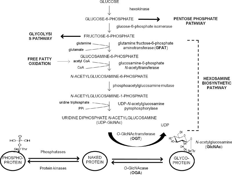
Diabetic retinopathy is a leading cause of blindness worldwide. Despite laser and surgical treatments, antiangiogenic and other therapies, and strict metabolic control, many patients progress to visual impairment and blindness. New insights are needed into the pathophysiology of diabetic retinopathy in order to develop new methods to improve the detection and treatment of disease and the prevention of blindness. Hyperglycemia and diabetes result in increased flux through the hexosamine biosynthetic pathway, which, in turn, results in increased PTM of Ser/Thr residues of proteins by O-linked β-N-acetylglucosamine (O-GlcNAc). O-GlcNAcylation is involved in regulation of many nuclear and cytoplasmic proteins in a manner similar to protein phosphorylation. Altered O-GlcNAc signaling has been implicated in the pathogenesis of diabetes and may play an important role in the pathogenesis of diabetic retinopathy. The goal of this review is to summarize the biology of the hexosamine biosynthesis pathway and O-GlcNAc signaling, to present the current evidence for the role of O-GlcNAc signaling in diabetes and diabetic retinopathy, and to discuss future directions for research on O-GlcNAc in the pathogenesis of diabetic retinopathy.
Keywords: Diabetes; Diabetic retinopathy; Glucose toxicity; Hexosamine biosynthesis pathway; O-GlcNAcylation.
© 2014 WILEY-VCH Verlag GmbH & Co. KGaA, Weinheim.
Figures

Similar articles
-
O-Linked β-N-acetylglucosamine (O-GlcNAc) modification: a new pathway to decode pathogenesis of diabetic retinopathy.Clin Sci (Lond). 2018 Jan 19;132(2):185-198. doi: 10.1042/CS20171454. Print 2018 Jan 31. Clin Sci (Lond). 2018. PMID: 29352075 Free PMC article. Review.
-
New insights: A role for O-GlcNAcylation in diabetic complications.Crit Rev Biochem Mol Biol. 2016 May-Jun;51(3):150-61. doi: 10.3109/10409238.2015.1135102. Epub 2016 Jan 24. Crit Rev Biochem Mol Biol. 2016. PMID: 26806492 Review.
-
Retinal O-linked N-acetylglucosamine protein modifications: implications for postnatal retinal vascularization and the pathogenesis of diabetic retinopathy.Mol Vis. 2013 May 21;19:1047-59. Print 2013. Mol Vis. 2013. PMID: 23734074 Free PMC article.
-
Protein O-GlcNAcylation coupled to Hippo signaling drives vascular dysfunction in diabetic retinopathy.Nat Commun. 2024 Oct 29;15(1):9334. doi: 10.1038/s41467-024-53601-x. Nat Commun. 2024. PMID: 39472558 Free PMC article.
-
O-GlcNAcylation under hypoxic conditions and its effects on the blood-retinal barrier in diabetic retinopathy.Int J Mol Med. 2014 Mar;33(3):624-32. doi: 10.3892/ijmm.2013.1597. Epub 2013 Dec 20. Int J Mol Med. 2014. PMID: 24366041
Cited by
-
FHL2 anchors mitochondria to actin and adapts mitochondrial dynamics to glucose supply.J Cell Biol. 2021 Oct 4;220(10):e201912077. doi: 10.1083/jcb.201912077. Epub 2021 Aug 3. J Cell Biol. 2021. PMID: 34342639 Free PMC article.
-
Galectins in the Pathogenesis of Common Retinal Disease.Front Pharmacol. 2021 May 17;12:687495. doi: 10.3389/fphar.2021.687495. eCollection 2021. Front Pharmacol. 2021. PMID: 34079467 Free PMC article. Review.
-
Topical Anti-ulcerogenic Effect of the Beta-adrenergic Blockers on Diabetic Foot Ulcers: Recent Advances and Future Prospectives.Curr Diabetes Rev. 2024;20(8):23-37. doi: 10.2174/0115733998249061231009093006. Curr Diabetes Rev. 2024. PMID: 37867269 Review.
-
RUNX1 can mediate the glucose and O-GlcNAc-driven proliferation and migration of human retinal microvascular endothelial cells.BMJ Open Diabetes Res Care. 2021 Aug;9(1):e001898. doi: 10.1136/bmjdrc-2020-001898. BMJ Open Diabetes Res Care. 2021. PMID: 34348917 Free PMC article.
-
Hyperglycemic Stress and Carbon Stress in Diabetic Glucotoxicity.Aging Dis. 2016 Jan 2;7(1):90-110. doi: 10.14336/AD.2015.0702. eCollection 2016 Jan. Aging Dis. 2016. PMID: 26816666 Free PMC article. Review.
References
-
- Yau JW, Roger SL, Kawasaki R, Lamoureux EL, Kowalski JW, Bek T, Chen SJ, Dekker JM, Fletcher A, Grauslund J, Haffner S, Hamman RF, Ikram MK, Kayama T, Klein BE, Klein R, Krishnaiah S, Mayurasakorn K, O'Hare JP, Orchard TJ, Porta M, Rema M, Roy MS, Sharma T, Mitchell P, Wong TY. Meta-Analysis for Eye Disease (META-EYE) Study Group, Global prevalence and major risk factors for diabetic retinopathy. Diabetes Care. 2012;35:556–564. - PMC - PubMed
-
- Antonetti DA, Klein R, Gardner TW. Diabetic retinopathy. N. Engl. J. Med. 2012;366:1227–1239. - PubMed
-
- Keech AC, Mitchell P, Summanen PA, O'Day J, Davis TM, Moffitt MS, Taskinen MR, Simes RJ, Tse D, Williamson E, Merrifield A, Laatikainen LT, d'Emden MC, O'Connell RL, Colman PG. FIELD Study Investigators, Effect of fenofibrate on the need for laser treatment for diabetic retinopathy (FIELD study): a randomised controlled trial. Lancet. 2007;370:1687–1697. - PubMed
-
- ACCORD Study Group ACCORD, Eye Study Group. Chew EY, Ambrosius WT, Davis MD, Danis RP, Gangaputra S, Greven CM, Hubbard L, Esser BA, Lovato JF, Perdue LH, Goff DC, Jr., Cushman WC, Ginsberg HN, Elam MB, Genuth S, Gerstein HC, Schubart U, Fine LJ. Effects of medical therapies on retinopathy progression in type 2 diabetes. N. Engl. J. Med. 2010;363:233–244. - PMC - PubMed
-
- Michaelides M, Kaines A, Hamilton RD, Fraser-Bell S, Rajendram R, Quhill F, Boos CJ, Xing W, Egan C, Peto T, Bunce C, Leslie RD, Hykin PG. A prospective randomized trial of intravitreal bevacizumab or laser therapy in the management of diabetic macular edema (BOLT study) 12-month data: report 2. Ophthalmology. 2010;117:1078–1086.e2. - PubMed
Publication types
MeSH terms
Substances
Grants and funding
LinkOut - more resources
Full Text Sources
Other Literature Sources
Medical
