. 2014 Jul;24(7):740-50.
doi: 10.1002/hipo.22264. Epub 2014 Mar 6.

Human hippocampal processing of environmental novelty during spatial navigation

Affiliations
Free PMC article

Human hippocampal processing of environmental novelty during spatial navigation

Raphael Kaplan et al. Hippocampus. 2014 Jul.
Free PMC article

Abstract

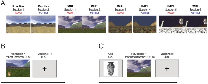
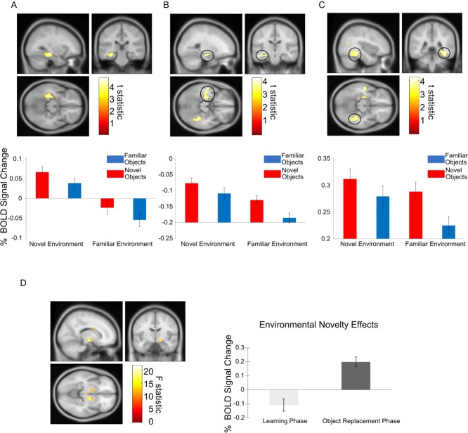
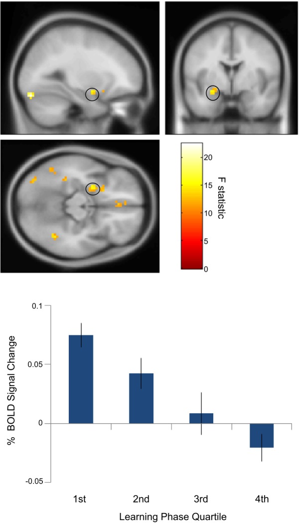
The detection and processing of novel information encountered as we explore our environment is crucial for learning and adaptive behavior. The human hippocampus has been strongly implicated in laboratory tests of novelty detection and episodic memory, but has been less well studied during more ethological tasks such as spatial navigation, typically used in animals. We examined fMRI BOLD activity as a function of environmental and object novelty as humans performed an object-location virtual navigation task. We found greater BOLD response to novel relative to familiar environments in the hippocampus and adjacent parahippocampal gyrus. Object novelty was associated with increased activity in the posterior parahippocampal/fusiform gyrus and anterior hippocampus extending into the amygdala and superior temporal sulcus. Importantly, whilst mid-posterior hippocampus was more sensitive to environmental novelty than object novelty, the anterior hippocampus responded similarly to both forms of novelty. Amygdala activity showed an increase for novel objects that decreased linearly over the learning phase. By investigating how participants learn and use different forms of information during spatial navigation, we found that medial temporal lobe (MTL) activity reflects both the novelty of the environment and of the objects located within it. This novelty processing is likely supported by distinct, but partially overlapping, sets of regions within the MTL.

Keywords: MTL; amygdala; content; context; fMRI.


Figures

Figure 1
Figure 1
Experimental Structure. A: Experimental environments shown from the participants' (first-person) perspective. Four different environments are presented in eight experimental sessions. The first two sessions (always the desert environments) provided practice outside of the scanner. Sessions 3–8 contained three novel-familiar environment repetitions with environment order, counterbalanced across participants. B: Learning phase trial structure. During learning trials, participants use a button box to navigate and “collect” three novel and three familiar (previously presented) objects (vase shown as example) four times each (a total of 24 trials per session). C: The object replacement phase, trial structure. After being cued for 3 s with a picture of an object that had been collected in the learning phase of the current session, participants were placed back in the environment and had to navigate to where they thought the object (object replacement) had been located during that learning period. [Color figure can be viewed in the online issue, which is available at wileyonlinelibrary.com.]
Figure 2
Figure 2
Environmental and object novelty during the navigation task. A: Left hippocampal activity corresponding to environmental novelty during navigation (above; peak voxel: x = −30; y = −28; z = −14; Z-score = 4.65; including both learning and object replacement phases). Percent signal change in a 10 mm sphere around the left hippocampal peak for all four conditions (navigating toward or replacing novel or familiar objects within novel or familiar environments, below, showing mean ± SEM over the 18 participants). B: Left anterior hippocampal activity, extending into the amygdala and superior temporal sulcus, corresponding to object novelty during navigation (above, peak voxel: x = 39; y = −40; z = −14; Z-score = 3.81). Percent signal change in a 10 mm sphere around the left anterior hippocampal peak for all four conditions (below, mean ± SEM). C: Left parahippocampal/fusiform activity corresponding to object novelty during navigation (above, peak voxel: x = 39; y = −40; z = −14; Z-score = 4.10). Percent signal change in a 10 mm sphere around the left parahippocampal/fusiform peak for all four conditions (mean ± SEM). D: Right ventral pallidum activity corresponding to the interaction between environmental novelty and experimental phase (left; peak voxel: x = 15; y = −10; z = −8; Z-score = 4.34; left ventral pallidum and midbrain/VTA effects visible in axial slice). Percent signal change in a 10 mm sphere around the right ventral pallidal peak during navigation in novel versus familiar environments during the learning and replacement phases (mean ± SEM). All activations are shown at the uncorrected threshold of P < 0.001 for display purposes and overlaid on the Montreal Neurological Institute 152 T1 image. [Color figure can be viewed in the online issue, which is available at wileyonlinelibrary.com.]
Figure 3
Figure 3
Temporal attenuation of amygdala object novelty effect. Above: Left amygdala activity (peak: x = −27; y = −1; z = −14; Z-score = 3.65) corresponding object novelty (novel versus familiar objects) showed a linear decrease over the course of the learning phase as relative novelty decreased. Below: Percent signal change extracted from a 10 mm sphere around the peak left amygdala voxel averaged across 18 participants for novel versus familiar objects for the first to fourth quartile of the learning phase. Activations are shown at the uncorrected threshold of P < 0.001 and overlaid on the Montreal Neurological Institute 152 T1 image for display purposes. [Color figure can be viewed in the online issue, which is available at wileyonlinelibrary.com.]

References

    1. Adolphs R. What does the amygdala contribute to social cognition? Ann NY Acad Sci. 2010;1191:42–61. - PMC - PubMed
    1. Balderston NL, Schultz DH, Helmstetter FJ. The human amygdala plays a stimulus specific role in the detection of novelty. Neuroimage. 2011;55:1889–1898. - PMC - PubMed
    1. Blackford JU, Buckholtz JW, Avery SN, Zald DH. A unique role for the human amygdala in novelty detection. Neuroimage. 2010;50:1188–1193. - PMC - PubMed
    1. Breiter HC, Etcoff NL, Whalen PJ, Kennedy WA, Rauch SL, Buckner RL, Strauss MM, Hyman SE, Rosen BR. Response and habituation of the human amygdala during visual processing of facial expression. Neuron. 1996;17:875–887. - PubMed
    1. Brett M, Anton JL, Valabregue R, Poline JB. Region of interest analysis using an SPM toolbox. Sendai: OHBM; 2002.

Publication types

LinkOut - more resources