. 2014 Apr;164(4):1930-51.
doi: 10.1104/pp.114.236117. Epub 2014 Feb 18.

Additional amphivasal bundles in pedicel pith exacerbate central fruit dominance and induce self-thinning of lateral fruitlets in apple

Affiliations

Additional amphivasal bundles in pedicel pith exacerbate central fruit dominance and induce self-thinning of lateral fruitlets in apple

Jean-Marc Celton et al. Plant Physiol. 2014 Apr.

Abstract

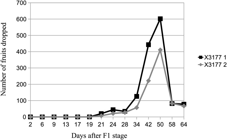
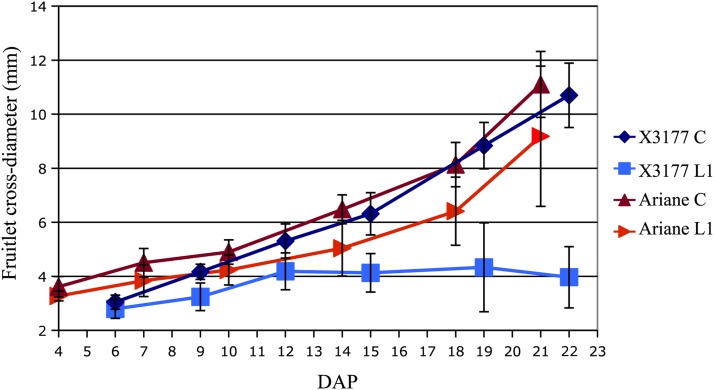
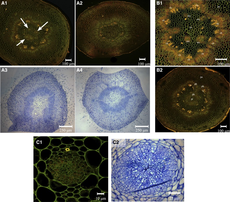
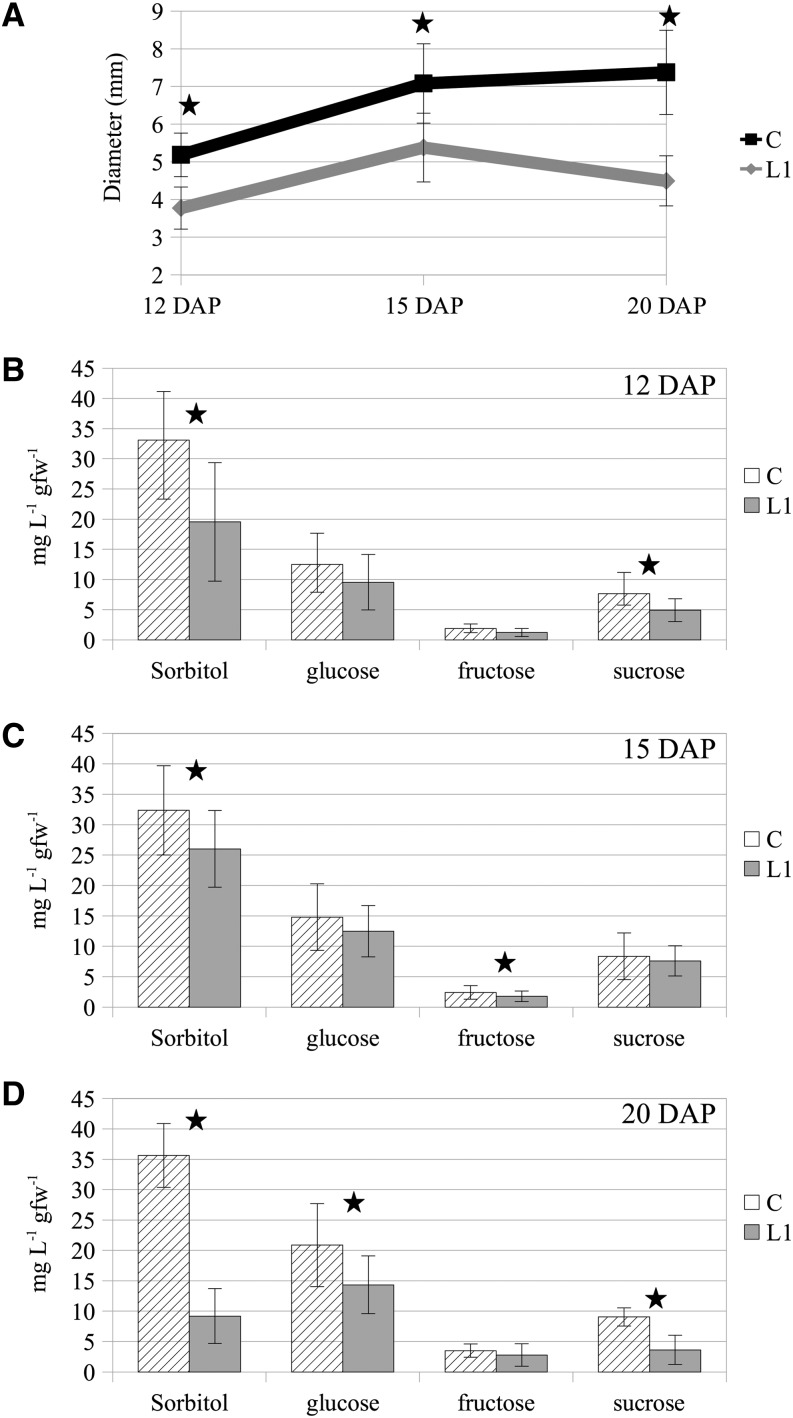
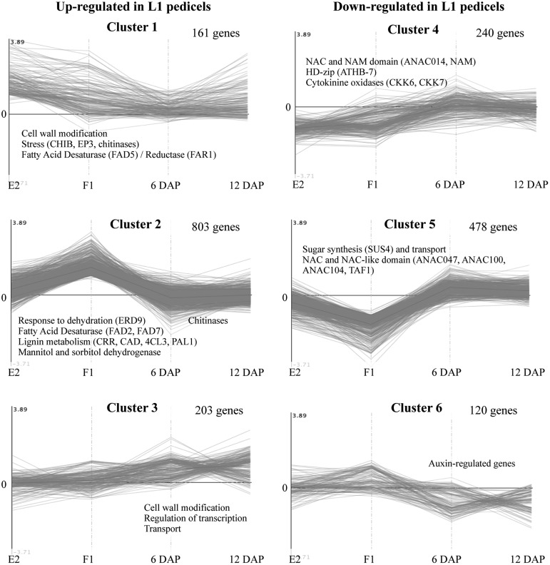
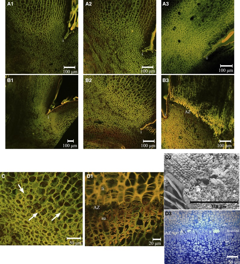
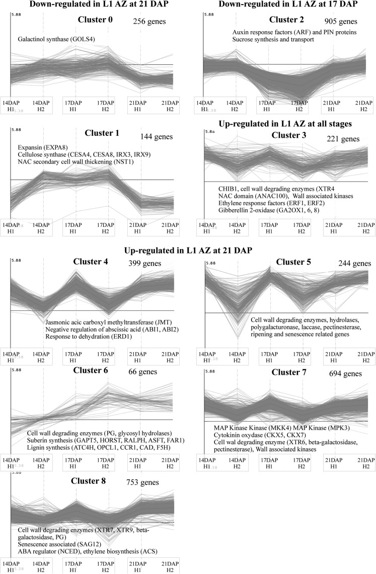
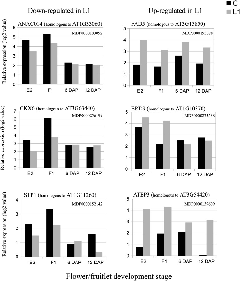
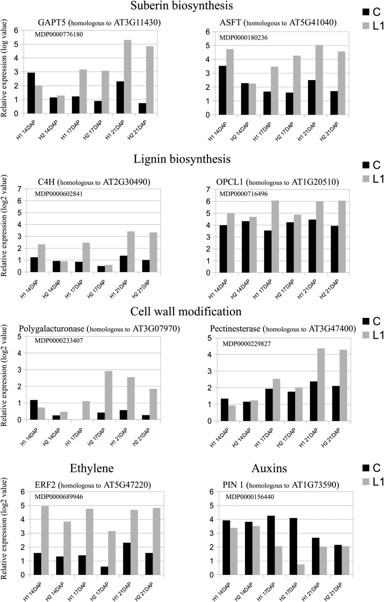
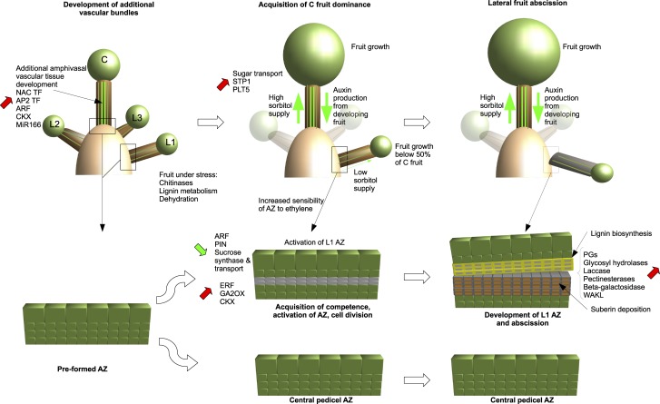
Apple (Malus × domestica) trees naturally produce an excess of fruitlets that negatively affect the commercial value of fruits brought to maturity and impact their capacity to develop flower buds the following season. Therefore, chemical thinning has become an important cultural practice, allowing the selective removal of unwanted fruitlets. As the public pressure to limit the use of chemical agents increases, the control of thinning becomes a major issue. Here, we characterized the self-thinning capacity of an apple hybrid genotype from the tree scale to the molecular level. Additional amphivasal vascular bundles were identified in the pith of pedicels supporting the fruitlets with the lowest abscission potential (central fruitlet), indicating that these bundles might have a role in the acquisition of dominance over lateral fruitlets. Sugar content analysis revealed that central fruitlets were better supplied in sorbitol than lateral fruitlets. Transcriptomic profiles allowed us to identify genes potentially involved in the overproduction of vascular tissues in central pedicels. In addition, histological and transcriptomic data permitted a detailed characterization of abscission zone development and the identification of key genes involved in this process. Our data confirm the major role of ethylene, auxin, and cell wall-remodeling enzymes in abscission zone formation. The shedding process in this hybrid appears to be triggered by a naturally exacerbated dominance of central fruitlets over lateral ones, brought about by an increased supply of sugars, possibly through additional amphivasal vascular bundles. The characterization of this genotype opens new perspectives for the selection of elite apple cultivars.


Figures

Figure 1.
Figure 1.
Fruit drop dynamics at the tree scale for both X3177 trees, expressed as the number of fruitlets dropped per tree following F1 stage (central flower opened).
Figure 2.
Figure 2.
Central (C) and L1 fruitlet mean cross-diameter kinetics of X3177 and cv Ariane trees in 2011, from 4 to 22 DAP. Error bars represent sd. [See online article for color version of this figure.]
Figure 3.
Figure 3.
Vascular patterns in central and L1 fruitlet stems of the apple hybrid X3177 and cv Ariane. A1 and A2, Pedicel cross section of X3177 central (A1) and L1 (A2) fruitlets 8 DAP, stained with acridine orange. Arrows indicate the positions of additional amphivasal bundles in the pith of central pedicels. A3 and A4, Pedicel cross section of cv Ariane central (A3) and L1 (A4) fruitlets 7 DAP, stained with toluidine blue. No amphivasal vascular bundles are observed in cv Ariane central pedicels. B1, Cross section of X3177 central flower pedicel at stage E2 (tight cluster, when the sepal lets the petals appear on central flower), stained with acridine orange. Surrounded by parenchyma (pa), vascular bundles (vb) are arranged in a ring pattern, with additional amphivasal bundles (am) located in the pith (pi). B2, Cross section of X3177 central pedicel cut at the base of the flower at stage E2, stained with acridine orange. Phloem (ph) tissues seem to abnormally penetrate into the pith of the pedicel. x, Xylem. C1, Cross section of amphivasal vascular bundle in the pith of the central pedicel of X3177 at the F1 stage (central flower open), stained with acridine orange. Cells are not yet organized, and only a single lignified cell is observed (in yellow). C2, Cross section of X3177 amphivasal vascular bundle in the pith of the central pedicel of X3177 19 DAP, stained with toluidine blue. Cells composing this bundle are organized in stratified layers, with compressed or sclerified cells at the center. The scale is represented by a bar at the bottom of each image.
Figure 4.
Figure 4.
A, L1 and central (C) diameters of selected hand-pollinated fruits at 12, 15, and 20 DAP of the apple hybrid X3177 in 2013. B to D, Sugar concentrations in central and L1 pedicels at three time points: 12 DAP (B), 15 DAP (C), and 20 DAP (D). Error bars represent sd. Statistically significant differences between the central and L1 pedicels as identified by Student’s t test are indicated by stars (P < 0.05). gfw, Grams fresh weight.
Figure 5.
Figure 5.
K-means clustering of significantly differentially expressed genes between central and L1 pedicels at four developmental stages (E2, F1, 6 DAP, and 12 DAP). The left column represents clusters of genes up-regulated in L1 at one or more developmental stages. The right column represents clusters of genes down-regulated in L1 at one or more developmental stages. The number of genes in each cluster is shown next to the cluster number. Names of selected genes or gene families are indicated in each cluster.
Figure 6.
Figure 6.
AZ development at the base of X3177 pedicels. A, Development of AZ in central pedicels at 14 DAP (A1), 17 DAP (A2), and 21 DAP (A3), stained with acridine orange. The AZ did not develop in the course of these three dates. i, Invagination. B, Development of AZ in L1 pedicels at 14 DAP (B1), 17 DAP (B2), and 21 DAP (B3), stained with acridine orange. The development of an AZ, represented by layers of different colored cells, is clear at 21 DAP. C, Closeup view of the AZ in L1 pedicels at 17 DAP, stained with acridine orange. Arrows indicate the positions of dividing cells. D, Closeup views of the AZ in L1 pedicels at 21 DAP. D1, Pedicel section stained with acridine orange. The AZ is divided in two; the top section (fruitlet side) is composed of large stratified cells with lignified cell walls (li), and the bottom section (bourse side) is composed of smaller stratified cells with possible suberin deposition on the inner face of primary cell walls (su). D2, Scanning electron microscopy of AZ in L1 pedicels at 21 DAP. Cell wall and middle lamella degradation is observed in the separation layer. D3, Pedicel stained with toluidine blue. The stratified cells composing the AZ and the separation layer can be observed. The scale is represented by a bar at the bottom of each image.
Figure 7.
Figure 7.
K-means clustering of significantly differentially expressed genes between central and L1 AZ at three developmental stages (14, 17, and 21 DAP) for the two X3177 biological replicates (H1 and H2). Clusters have been regrouped according to their profiles. The number of genes in each cluster is shown next to the cluster number. Names of selected genes or gene families are indicated in each cluster.
Figure 8.
Figure 8.
Kinetics of the expression values of genes differentially expressed between central (C) and L1 X3177 apple pedicels as determined by microarray analysis at four developmental stages (E2, F1, 6 DAP, and 12 DAP). The left column shows expression values (log values) of genes significantly down-regulated in L1 compared with central pedicel sections. The right column shows expression values (log values) of genes significantly up-regulated in L1 compared with central pedicel sections. The array data were normalized with the lowess method. Normalized intensities (i.e. expression levels) were then subtracted from the background.
Figure 9.
Figure 9.
Kinetics of the expression values of genes differentially expressed between central (C) and L1 AZs of the two X3177 apple biological replicates (H1 and H2) as determined by microarray analysis at three developmental stages (14, 17, and 21 DAP). The kinetics of expression values of selected genes involved in suberin biosynthesis, lignin biosynthesis, cell wall modification, ethylene response factor, and auxin transport are represented. The transcript levels were normalized with the lowess method. Intensity values were then subtracted from the background.
Figure 10.
Figure 10.
Model of the development of apple fruitlets and their respective AZs. This model was developed from the analyses of X3177 corymbs, pedicel vascular tissue, and AZ development. Data presented from analyses in this article are schematically summarized in relation to the relative positions of the fruitlets within the corymb. In this model, additional bundles develop early in the central fruit pedicel and may provide it with additional resources such as sorbitol, allowing it to develop a strong dominance over the lateral ones. Global gene expression data indicate a general trend toward an up-regulation of genes involved in vascular tissue differentiation in central pedicels together with a down-regulation of genes involved in sugar transport in L1 pedicels. AZ activation is triggered 4 to 5 d following lateral fruit growth arrest and is initiated with a cycle of cell divisions. Following the formation of layers of isodiametrically flattened cells, we observe a degradation of cell walls and middle lamella at the separation zone by multiple cell wall-modifying enzymes. Cells located on the pedicel side become impregnated with lignin, while cells located on the bourse side accumulate suberin depositions. C, Central fruitlet; L1, small lateral fruitlet; L2, medium lateral fruitlet; L3, big lateral fruitlet.

References

    1. Abot A (2010) Caractérisation des fibres longues de chanvre (Cannabis sativa) en vue de leurs utilisations dans des matériaux composites. PhD thesis. Université de Poitier, Poitier, France
    1. Agustí J, Merelo P, Cercós M, Tadeo FR, Talón M. (2008) Ethylene-induced differential gene expression during abscission of citrus leaves. J Exp Bot 59: 2717–2733 - PMC - PubMed
    1. Agustí J, Merelo P, Cercós M, Tadeo FR, Talón M. (2009) Comparative transcriptional survey between laser-microdissected cells from laminar abscission zone and petiolar cortical tissue during ethylene-promoted abscission in citrus leaves. BMC Plant Biol 9: 127. - PMC - PubMed
    1. Aida M, Ishida T, Tasaka M. (1999) Shoot apical meristem and cotyledon formation during Arabidopsis embryogenesis: interaction among the CUP-SHAPED COTYLEDON and SHOOT MERISTEMLESS genes. Development 126: 1563–1570 - PubMed
    1. Alferez F, Zhong GY, Burns JK. (2007) A citrus abscission agent induces anoxia- and senescence-related gene expression in Arabidopsis. J Exp Bot 58: 2451–2462 - PubMed

Publication types

MeSH terms