. 2014 Feb 18;111(7):2494-9.
doi: 10.1073/pnas.1318962111. Epub 2014 Feb 3.

The tripartite motif coiled-coil is an elongated antiparallel hairpin dimer

Affiliations

The tripartite motif coiled-coil is an elongated antiparallel hairpin dimer

Jacint G Sanchez et al. Proc Natl Acad Sci U S A. .

Abstract

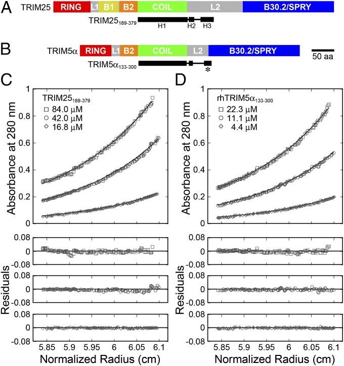
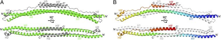
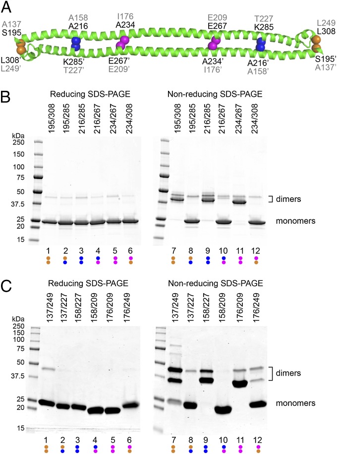
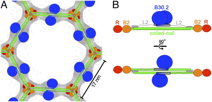
Tripartite motif (TRIM) proteins make up a large family of coiled-coil-containing RING E3 ligases that function in many cellular processes, particularly innate antiviral response pathways. Both dimerization and higher-order assembly are important elements of TRIM protein function, but the atomic details of TRIM tertiary and quaternary structure have not been fully understood. Here, we present crystallographic and biochemical analyses of the TRIM coiled-coil and show that TRIM proteins dimerize by forming interdigitating antiparallel helical hairpins that position the N-terminal catalytic RING domains at opposite ends of the dimer and the C-terminal substrate-binding domains at the center. The dimer core comprises an antiparallel coiled-coil with a distinctive, symmetric pattern of flanking heptad and central hendecad repeats that appear to be conserved across the entire TRIM family. Our studies reveal how the coiled-coil organizes TRIM25 to polyubiquitylate the RIG-I/viral RNA recognition complex and how dimers of the TRIM5α protein are arranged within hexagonal arrays that recognize the HIV-1 capsid lattice and restrict retroviral replication.

Keywords: X-ray crystallography; antiparallel dimer; disulfide crosslinking.


Conflict of interest statement

The authors declare no conflict of interest.

Figures

Fig. 1.
Fig. 1.
Domain organization and dimerization of TRIM proteins. (A) Schematic of the domain structure of TRIM25. The principal domains and linker regions are RING (red), L1 (gray), B-box 1 (yellow), B-box 2 (orange), coiled-coil (green), L2 (gray), and B30.2/SPRY (blue). The TRIM25189–379 construct used in this study is shown beneath (black), with the secondary structure derived from the crystal structure (rectangles represent helices). (B) Analogous schematic of the domain structure of TRIM5α. TRIM5α does not contain a B-box 1 domain and has a shorter L2. The TRIM5α133–300 construct used in this study is shown beneath (black). The asterisk denotes a predicted helix (H3) that crosses from L2 into the B30.2 domain. (C) TRIM25189–379 is a stable dimer in solution. Equilibrium sedimentation distributions of the indicated protein concentrations are shown for the rotor speed of 12,000 rpm. (Upper) Absorbance measurements (open symbols; 280 nm) and best-fit curves (solid lines). (Lower) Residual differences. Equilibrium distributions were also measured at rotor speeds of 17,000 and 23,000 rpm (not shown for clarity), and all of the data were globally fit to a single-species model in which the molecular weight (Mobs) was allowed to float (Mobs = 41,674 Da; Mcalc = 21,835 Da; Mobs/Mcalc = 1.91). Fits in which the molecular weight was fixed to that of a dimer are shown in Fig. S1A. (D) TRIM5α133–300 is also a stable dimer in solution (Mobs = 43,505 Da; Mcalc = 23,038 Da; Mobs/Mcalc = 1.89). See Fig. S1D for fits to a single-species model with a fixed dimer molecular weight.
Fig. 2.
Fig. 2.
Structure of the TRIM25189–379 dimer. (A) Orthogonal views of the dimer in ribbons representation, with the coiled-coil and L2 segments colored in green and gray, respectively (matching the color scheme of Fig. 1). (B) Orthogonal views of the dimer with one subunit colored in rainbow gradient, with blue at the N terminus and red at the C terminus, and the other subunit in white.
Fig. 3.
Fig. 3.
Dimeric packing of TRIM coiled-coil helices. (A) Coiled-coil formed by the H1 and H1′ helices in the TRIM25189–379 structure. Side-chains that mediate interhelix packing interactions are numbered and shown as spheres, with the a, d, and h positions colored in red, yellow, and cyan, respectively. Circled numbers indicate mutation sites analyzed in C. The dimer symmetry axis (black oval) runs perpendicular to the page. (B) Expanded view of the central region boxed in A. Side-chains that mediate important packing interactions are shown as spheres, colored as in A and labeled, as are buried water molecules (orange). (C) Thermofluor melting curves of wild-type (filled circles), L252A (open circles), M209A (filled diamonds), and V223A (filled squares). The high fluorescence signal for L252A at 25 °C indicates that the hydrophobic residues of this mutant are already exposed. Error bars represent the SDs from 4 replicates performed in parallel. (D) Structure-to-sequence alignment. The graph shows a multiple sequence alignment of 54 different human TRIM coiled-coil/L2 sequences displayed in logo format (see Fig. S4 for full alignment). The sequence alignment is overlaid with percentage buried surface area plots calculated using the entire TRIM25189–379 structure (light gray bars) or the H1/H1′ helices only (dark gray bars). Heptad/hendecad residue assignments are color coded as in A. The TRIM25 sequence is shown at the top and the aligned TRIM5α sequence at the bottom, with the first and last residue numbers indicated.
Fig. 4.
Fig. 4.
Disulfide crosslinking of TRIM25 and TRIM5α dimers. (A) H1 and H2 regions of the TRIM25189–379 structure showing positions of residue pairs chosen for cysteine mutagenesis. Equivalent TRIM25 and TRIM5α residues are labeled in black and gray, respectively. (B) Electrophoretic profiles of purified TRIM25189–379 double-cysteine mutants that were dialyzed under nonreducing conditions, then denatured in SDS-PAGE buffer under reducing (Left) or nonreducing (Right) conditions. Molecular weight marker positions are labeled on the left. Positions of monomers and crosslinked dimers are labeled on the right. Note that the symmetric dimer is expected to produce two types of intermolecular disulfide-crosslinked species: one in which both cysteine pairs are oxidized (lower bands) and another in which one of the pairs is reduced (upper bands). Data are representative of 3 independent experiments. (C) Profiles of rhesus TRIM5α133–300 cysteine mutants that were dialyzed under mildly reducing conditions and then prepared for SDS-PAGE, as described for TRIM25189–379. Data are representative of 3 independent experiments.
Fig. 5.
Fig. 5.
Models of quaternary TRIM5α interactions. (A) Schematic model of the TRIM5α hexagonal lattice, showing the deduced positions of the different domains and overlaid with the cryoEM projection map (gray contours) (18). Domains are colored as in Fig. 1B. (B) Schematic model of the full-length TRIM5α dimer. The C-terminal B30.2 domains (blue) are shown packed against one side of the coiled-coil domain via a putative extended H3 helix (colored in gray to blue gradient and outlined in black) that spans both L2 and B30.2 sequences and forms a 4-helix bundle with the coiled-coil, as seen in the TRIM25 structure.

References

    1. Han K, Lou DI, Sawyer SL. Identification of a genomic reservoir for new TRIM genes in primate genomes. PLoS Genet. 2011;7(12):e1002388. - PMC - PubMed
    1. McNab FW, Rajsbaum R, Stoye JP, O’Garra A. Tripartite-motif proteins and innate immune regulation. Curr Opin Immunol. 2011;23(1):46–56. - PubMed
    1. Gack MU, et al. TRIM25 RING-finger E3 ubiquitin ligase is essential for RIG-I-mediated antiviral activity. Nature. 2007;446(7138):916–920. - PubMed
    1. Oshiumi H, Matsumoto M, Seya T. Ubiquitin-mediated modulation of the cytoplasmic viral RNA sensor RIG-I. J Biochem. 2012;151(1):5–11. - PubMed
    1. Stremlau M, et al. The cytoplasmic body component TRIM5α restricts HIV-1 infection in Old World monkeys. Nature. 2004;427(6977):848–853. - PubMed

Publication types

Associated data