. 2014 Feb 18;111(7):2548-53.
doi: 10.1073/pnas.1324297111. Epub 2014 Feb 3.

Preleukemic mutations in human acute myeloid leukemia affect epigenetic regulators and persist in remission

Affiliations

Preleukemic mutations in human acute myeloid leukemia affect epigenetic regulators and persist in remission

M Ryan Corces-Zimmerman et al. Proc Natl Acad Sci U S A. .

Abstract

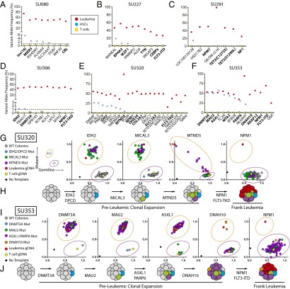
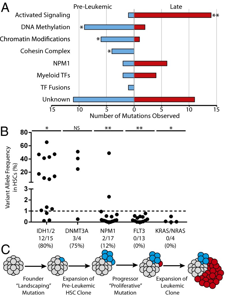
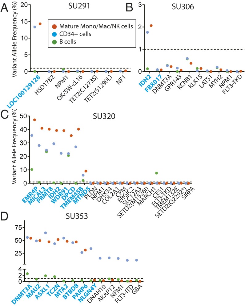
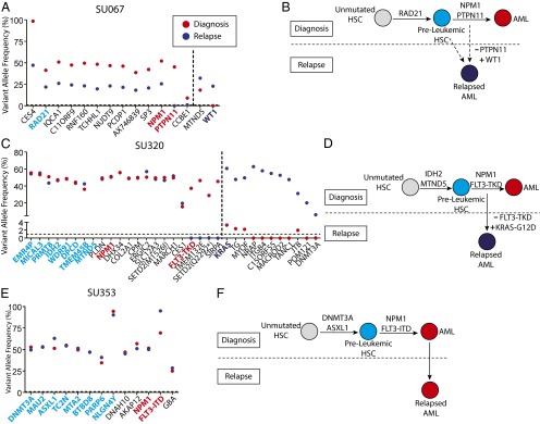
Cancer is widely characterized by the sequential acquisition of genetic lesions in a single lineage of cells. Our previous studies have shown that, in acute myeloid leukemia (AML), mutation acquisition occurs in functionally normal hematopoietic stem cells (HSCs). These preleukemic HSCs harbor some, but not all, of the mutations found in the leukemic cells. We report here the identification of patterns of mutation acquisition in human AML. Our findings support a model in which mutations in "landscaping" genes, involved in global chromatin changes such as DNA methylation, histone modification, and chromatin looping, occur early in the evolution of AML, whereas mutations in "proliferative" genes occur late. Additionally, we analyze the persistence of preleukemic mutations in patients in remission and find CD34+ progenitor cells and various mature cells that harbor preleukemic mutations. These findings indicate that preleukemic HSCs can survive induction chemotherapy, identifying these cells as a reservoir for the reevolution of relapsed disease. Finally, through the study of several cases of relapsed AML, we demonstrate various evolutionary patterns for the generation of relapsed disease and show that some of these patterns are consistent with involvement of preleukemic HSCs. These findings provide key insights into the monitoring of minimal residual disease and the identification of therapeutic targets in human AML.

Keywords: clonal evolution; preleukemia.


Conflict of interest statement

The authors declare no conflict of interest.

Figures

Fig. 1.
Fig. 1.
Sequencing and single-cell genotyping assays identify preleukemic mutations and HSCs in a diverse cohort of human AML patients. (A–F) Targeted amplicon sequencing was performed on all leukemic mutations identified by exome sequencing in six AML patients. Targeted sequencing was performed on gDNA isolated from FACS-purified leukemia cells (red, CD99+ TIM3+), T cells (yellow, CD3+), and HSCs (blue, CD34+/CD38−/CD99−/TIM3−) for each patient. The dotted line represents the threshold of detection. Gene names highlighted in bold indicate recurrently mutated genes in AML. (G and I) Single HSC-derived colony genotyping assays were performed for patient samples SU320 (G) and SU353 (I) using custom TaqMan SNP genotyping assays specific for all recurrent mutations. Each colony was classified as wild type or mutant for each mutation based on comparison with leukemia (red), T-cell (yellow), or no template (black) control reactions. (H and J) The single-cell data from G and I were used to diagram the evolutionary history of cases SU320 (H) and SU353 (J), where preleukemic mutations are sequentially acquired followed by late mutations never found in colonies derived from HSCs.
Fig. 2.
Fig. 2.
Mutation acquisition in AML occurs in patterns with preleukemic landscaping mutations followed by late proliferative mutations. (A) Exome sequencing and targeted amplicon sequencing data identifying preleukemic and late mutations from 16 patients (Table S1) were pooled. Recurrent mutations (74 total) were stratified according to the classification system established by the TCGA, and assigned to be preleukemic or late based on detection in HSCs as described in Table S4. The number of mutations in each category is indicated on the x axis. (B) Additional AML cases were selected for the presence of recurrent mutations in IDH1/IDH2 (n = 15), DNMT3A (n = 4), NPM1 (n = 17), FLT3 (n = 13), or KRAS/NRAS (n = 4). Each dot represents one case (see Table S2 for patient information). The dotted line represents the threshold of detection. The ratio of cases with preleukemic mutation of a given gene to the total number of cases with that mutation analyzed is shown below the plot. (C) A model for the acquisition of mutations in AML. (**P < 0.001, *P < 0.05; NS, not significant, χ2 test).
Fig. 3.
Fig. 3.
Preleukemic mutations and HSCs persist in remission. (A–D) Bone marrow mononuclear cells from patient remission samples were sorted for CD19+ B cells, CD14+/CD11b+/CD56+ mature monocytes, macrophages, and NK cells, and CD34+ hematopoietic progenitors. Targeted amplicon sequencing was performed for all leukemic mutations in all sorted populations. The dotted line represents the threshold of detection. All mutations identified as being preleukemic at diagnosis are highlighted in blue.
Fig. 4.
Fig. 4.
Preleukemic mutations are retained at relapse. (A, C, and E) Exome sequencing was performed to identify mutations detectable at relapse that were not detected at diagnosis. Targeted amplicon sequencing of all diagnosis-specific and relapse-specific mutations was performed in FACS-purified leukemic cells (CD99+ TIM3+) from the disease at diagnosis (red) and at relapse (dark blue). Diagnosis-specific mutations are depicted to the left of the dotted line, whereas relapse-specific mutations are depicted to the right. All mutations identified as being preleukemic at diagnosis are highlighted in light blue, whereas recurrent mutations identified as being late mutations at diagnosis are highlighted in red. Recurrent mutations identified at relapse are highlighted in dark blue. (B, D, and F) Evolutionary models for the progression of disease in each case are presented, with depiction of recurrently mutated genes in each model. The solid arrows represent determined evolutionary steps, and the dotted arrows represent inferred evolutionary steps.

Comment in

  • On the origin of leukemic species.
    Vasanthakumar A, Godley LA. Vasanthakumar A, et al. Cell Stem Cell. 2014 Apr 3;14(4):421-2. doi: 10.1016/j.stem.2014.03.008. Cell Stem Cell. 2014. PMID: 24702991

References

    1. Estey E, Döhner H. Acute myeloid leukaemia. Lancet. 2006;368(9550):1894–1907. - PubMed
    1. Löwenberg B, Downing JR, Burnett A. Acute myeloid leukemia. N Engl J Med. 1999;341(14):1051–1062. - PubMed
    1. Ley TJ, et al. DNMT3A mutations in acute myeloid leukemia. N Engl J Med. 2010;363(25):2424–2433. - PMC - PubMed
    1. Welch JS, et al. The origin and evolution of mutations in acute myeloid leukemia. Cell. 2012;150(2):264–278. - PMC - PubMed
    1. Ley TJ, et al. DNA sequencing of a cytogenetically normal acute myeloid leukaemia genome. Nature. 2008;456(7218):66–72. - PMC - PubMed

Publication types