. 2014 Feb 18;111(7):2590-5.
doi: 10.1073/pnas.1400744111. Epub 2014 Feb 3.

Cartilage-specific deletion of Mig-6 results in osteoarthritis-like disorder with excessive articular chondrocyte proliferation

Affiliations

Cartilage-specific deletion of Mig-6 results in osteoarthritis-like disorder with excessive articular chondrocyte proliferation

Ben Staal et al. Proc Natl Acad Sci U S A. .

Abstract

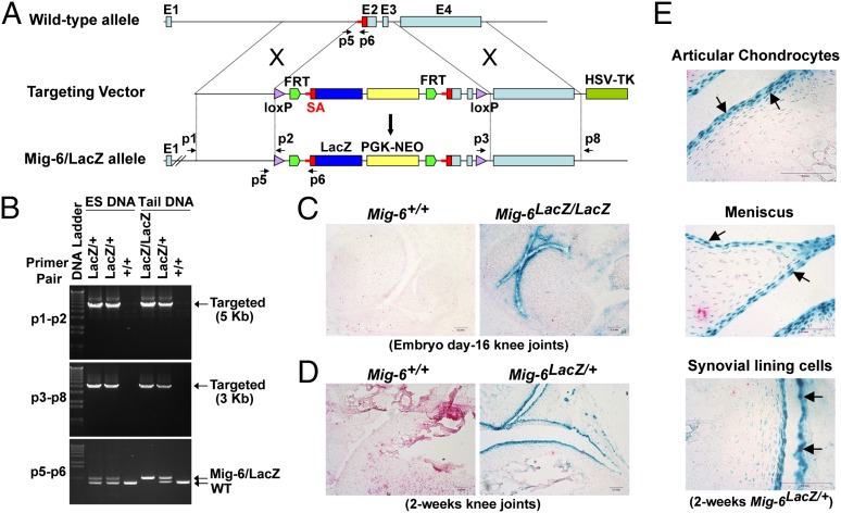
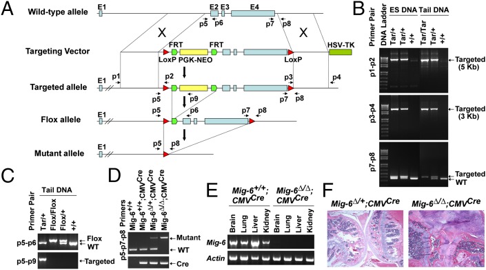
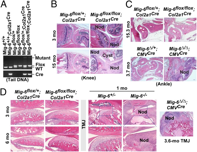
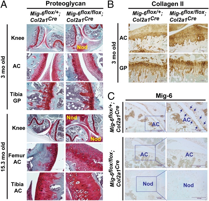
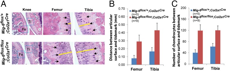
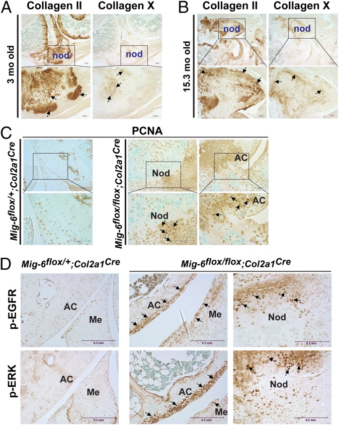
A deficiency of mitogen-inducible gene-6 (Mig-6) in mice leads to the development of an early-onset, osteoarthritis (OA)-like disorder in multiple synovial joints, underlying its importance in maintaining joint homeostasis. Here we determined what joint tissues Mig-6 is expressed in and what role chondrocytes play in the Mig-6-deficient OA-like disorder. A Mig-6/lacZ reporter mouse strain expressing β-galactosidase under the control of the Mig-6 gene promoter was generated to determine Mig-6 expression in joint tissues. By β-galactosidase staining, we demonstrated that Mig-6 was uniquely expressed in the cells across the entire surface of the synovial joint cavity, including chondrocytes in the superficial zone of articular cartilage and in the meniscus, as well as synovial lining cells. By crossing Mig-6-floxed mice to Col2a1-Cre transgenic mice, to generate cartilage-specific deletion of Mig-6, we demonstrated that deficiency of Mig-6 in the chondrocytes results in a joint phenotype that only partially recapitulates the OA-like disorder of the Mig-6-deficient mice: Ubiquitous deletion of Mig-6 led to the OA-like disorder in multiple joints, whereas cartilage-specific deletion affected the knees but rarely other joints. Furthermore, chondrocytes with Mig-6 deficiency showed excessive proliferative activities along with enhanced EGF receptor signaling in the articular cartilage and in the abnormally formed osteophytes. Our findings provide insight into the crucial requirement for Mig-6 in maintaining joint homeostasis and in regulating chondrocyte activities in the synovial joints. Our data also suggest that other cell types are required for fully developing the Mig-6-deficient OA-like disorder.

Keywords: EGFR; Errfi1; Gene 33; RALT.


Conflict of interest statement

The authors declare no conflict of interest.

Figures

Fig. 1.
Fig. 1.
Generation of Mig-6/lacZ reporter mice and determination of Mig-6–expressing cells in synovial joints. (A) The strategy for constructing the targeting vector to generate Mig-6/lacZ reporter mice. (B) Allele-specific PCR confirmation of the Mig-6/lacZ alleles (lacZ/+ and lacZ/lacZ) in the ES clones and the resulting mice. The p1-p2 and p3-p8 primer pairs amplified a 5-kb and a 3-kb DNA fragment, respectively, both specific for the Mig-6/lacZ alleles after proper homologous recombination. The p5–p6 primers were used for PCR genotyping to distinguish the wild-type (WT) and Mig-6/lacZ alleles. (C) β-gal staining of the day-16 embryonic Mig-6+/+ and Mig-6lacZ/lacZ knee joints. (D) β-gal staining of 2-wk-old Mig-6+/+ and Mig-6lacZ/+ knee joints. High lacZ reporter activity was detected across the entire surface of the joint cavity. (E) High-magnification images of Mig-6–expressing cells including the articular chondrocytes, chondrocytes in the meniscus, and the synovial lining cells (arrows) in a 2-wk-old Mig-6lacZ/+ knee joint.
Fig. 2.
Fig. 2.
Generation and characterization of Mig-6–floxed mice. (A) The strategy for the generation of Mig-6–floxed mice. (B) Confirmation of proper homologous recombination by allele-specific PCR. Genomic DNA from ES clones and the resulting mice was used for PCR amplification with primer pairs (p1–p2 and p3–p4) specific for the targeted alleles (Tar/+ and Tar/Tar). The p7–p8 primer pair was used for distinguishing between WT and the targeted allele. (C) PCR confirmation of the Mig-6flox allele using mouse tail DNA. The p5–p9 primers specifically amplified the targeted allele, whereas the p5–p6 pair separated the WT and floxed alleles. (D) Deletion of Mig-6 in Mig-6∆/∆;CMVCre mice by crossing Mig-6–floxed mice to CMV-cre mice. The p5–p8 primers detected the mutant alleles (Mig-6∆/∆ and Mig-6∆/+), and the p7–p8 pair detected the WT allele. The presence of CMV-Cre was determined by Cre-specific PCR. (E) RT-PCR detection of Mig-6 expression in the tissues of the Mig-6+/+;CMVCre and Mig-6∆/∆;CMVCre mice. Actin was used as an internal control. (F) Knee joints from 3.6-mo-old Mig-6∆/+;CMVCre and Mig-6∆/∆;CMVCre mice; Mig-6∆/∆;CMVCre mice developed OA-like disorder, phenocopying Mig-6−/− mice.
Fig. 3.
Fig. 3.
Cartilage-specific deletion of Mig-6 results in OA-like phenotype in the knee joints. (A) PCR genotyping of tail DNA from a Mig-6flox/flox;Col2a1Cre mouse and from control mice using the p5–p7–p8 triple primer combination. The p5–p8 primers detected the mutant allele, whereas the p7-p8 primers detected and distinguished floxed and WT alleles. *Note: Only a small population of cells in the tails expresses Col2a1 and thus Cre recombinase; floxed alleles were unmodified in the cells without Cre expression, resulting in PCR detection of a weak signal for the mutant allele together with a strong signal for the flox allele in the Mig-6flox/flox;Col2a1Cre tail. (B) Knee joints from Mig-6flox/+;Col2a1Cre mice and Mig-6flox/flox;Col2a1Cre mice at the indicated ages. (C) Ankles from Mig-6flox/+;Col2a1Cre and Mig-6flox/flox;Col2a1Cre mice at 15.3 mo, and from Mig-6∆/+;CMVCre and Mig-6∆/∆;CMVCre mice at 3.7 mo. (D) TMJs from Mig-6flox/+;Col2a1Cre and Mig-6flox/flox;Col2a1Cre mice at 3 and 6 mo, from Mig-6+/− and Mig-6−/− mice at 1 mo, and from a Mig-6∆/∆;CMVCre mouse at 3.6 mo. Osteophytes are indicated by “Nod” and subchondral cysts by “Cyst”.
Fig. 4.
Fig. 4.
Proteoglycan and collagen II distributions and detection of Mig-6 expression in the knee joints of Mig-6flox/flox;Col2a1Cre and the control mice. (A) Safranin O staining was used to detect proteoglycan in the knee joints of the Mig-6flox/+;Col2a1Cre and Mig-6flox/flox;Col2a1Cre mice at 3 and 15.3 mo of age. The osteophytes (labeled “nod”) in the Mig-6flox/flox;Col2a1Cre joints at both ages produced an abundance of proteoglycans. The thickness of the proteoglycan-positive articular cartilage (AC) in both the femur and tibia was significantly increased in the Mig-6flox/flox;Col2a1Cre joints especially in older mice, relative to those of Mig-6flox/+;Col2a1Crecontrol joints. The distribution of proteoglycans in the growth plates (GP) of both strains appeared similar. (B) IHC staining of collagen II in the articular cartilage and growth plate of 3-mo-old Mig-6flox/+;Col2a1Cre and Mig-6flox/flox;Col2a1Cre knee joints. (C) Detection of Mig-6 expression in the Mig-6flox/+;Col2a1Cre and Mig-6flox/flox;Col2a1Cre knee joints by IHC staining. The arrows indicate representative positively stained cells.
Fig. 5.
Fig. 5.
Chondrocyte-specific deletion of Mig-6 resulted in thickening of articular cartilage accompanying increase of chondrocyte numbers. (A) Representative knee joints of the Mig-6flox/+;Col2a1Cre and Mig-6flox/flox;Col2a1Cre mice (age 16.1 mo) are shown. The arrows indicate the tidemarks, and the yellow bars measure the distance between the tidemark and the surface of the articular cartilage. (B) The distance between the articular surface and the tidemark is significantly increased in both femurs and tibias of Mig-6flox/flox;Col2a1Cre mice. (C) The number of chondrocytes in the articular cartilage between articular surface and tidemark (40× magnification fields) was also significantly increased in the Mig-6flox/flox;Col2a1Cre mice relative to their control mice (ages between 13.9 and 16.1 mo). The rectangles indicate the average values, and the error bars indicate the SD.
Fig. 6.
Fig. 6.
Detection of collagens type II, type X, PCNA, p-EGFR, and p-ERK in the Mig-6flox/flox;Col2a1Cre knee joint. Strong staining of collagen II was detected within the osteophytes (labeled “nod”) in both 3-mo-old (A) and 15.3-mo-old (B) knee joints. In the inner zones of the osteophytes were collagen X–positive hypertrophic chondrocytes surrounded by collagen II–positive chondrocytes. (C) Detection of proliferating cells in the osteophytes and articular cartilage by IHC staining of PCNA in knee joint sections derived from Mig-6flox/+;Col2a1Cre and Mig-6flox/flox;Col2a1Cre mice at 3 mo of age. The PCNA-positive cells were detected in osteophytes and around the articular cartilage (AC) in the Mig-6flox/flox;Col2a1Cre knee. (D) Detection of p-EGFR and p-ERK in the Mig-6flox/+;Col2a1Cre and Mig-6flox/flox;Col2a1Cre knee joints by IHC staining. The arrows indicate representative positively stained (brown) cells.

References

    1. Zhang YW, Vande Woude GF. Mig-6, signal transduction, stress response and cancer. Cell Cycle. 2007;6(5):507–513. - PubMed
    1. Zhang YW, Vande Woude GF. MIG-6 and SPRY2 in the regulation of receptor tyrosine kinase signaling: Balancing act via negative feedback loops. In: Cheng Y, editor. Future Aspects of Tumor Suppressor Gene. Rijeka, Croatia: InTech; 2013. pp. 199–221.
    1. Anastasi S, et al. Feedback inhibition by RALT controls signal output by the ErbB network. Oncogene. 2003;22(27):4221–4234. - PubMed
    1. Zhang X, et al. Inhibition of the EGF receptor by binding of MIG6 to an activating kinase domain interface. Nature. 2007;450(7170):741–744. - PMC - PubMed
    1. Frosi Y, et al. A two-tiered mechanism of EGFR inhibition by RALT/MIG6 via kinase suppression and receptor degradation. J Cell Biol. 2010;189(3):557–571. - PMC - PubMed

Publication types

MeSH terms

Substances