. 2014 Feb 18;111(7):2722-7.
doi: 10.1073/pnas.1317454111. Epub 2014 Jan 13.

Leukocyte immunoglobulin-like receptor B1 is critical for antibody-dependent dengue

Affiliations

Leukocyte immunoglobulin-like receptor B1 is critical for antibody-dependent dengue

Kuan Rong Chan et al. Proc Natl Acad Sci U S A. .

Abstract

Viruses must evade the host innate defenses for replication and dengue is no exception. During secondary infection with a heterologous dengue virus (DENV) serotype, DENV is opsonized with sub- or nonneutralizing antibodies that enhance infection of monocytes, macrophages, and dendritic cells via the Fc-gamma receptor (FcγR), a process termed antibody-dependent enhancement of DENV infection. However, this enhancement of DENV infection is curious as cross-linking of activating FcγRs signals an early antiviral response by inducing the type-I IFN-stimulated genes (ISGs). Entry through activating FcγR would thus place DENV in an intracellular environment unfavorable for enhanced replication. Here we demonstrate that, to escape this antiviral response, antibody-opsonized DENV coligates leukocyte Ig-like receptor-B1 (LILRB1) to inhibit FcγR signaling for ISG expression. This immunoreceptor tyrosine-based inhibition motif-bearing receptor recruits Src homology phosphatase-1 to dephosphorylate spleen tyrosine kinase (Syk). As Syk is a key intermediate of FcγR signaling, LILRB1 coligation resulted in reduced ISG expression for enhanced DENV replication. Our findings suggest a unique mechanism for DENV to evade an early antiviral response for enhanced infection.

Keywords: early innate immune response; immune evasion; innate immune signaling.


Conflict of interest statement

The authors declare no conflict of interest.

Figures

Fig. 1.
Fig. 1.
ADE differs in THP-1 subclones. (A) Percentage of internalized DiD-labeled DENV-2 30 min postinfection under DENV-2 or ADE conditions in THP-1, THP-1.2R, and THP-1.2S. (B) Plaque titers of THP-1, THP-1.2R, or THP-1.2S when infected with DENV-2 opsonized with different h3H5 concentrations 72 h postinfection (hpi). Dotted lines indicate plaque titers following DENV-2–only infection, with no significant differences observed between the cell lines. (C) Time course of viral RNA copy numbers in THP-1.2R or THP-1.2S under ADE conditions. (D) Heat map showing fold change of ISG expression in THP-1.2R and THP-1.2S at 6 hpi under ADE conditions. (E) Fold change in transcript levels of interferons in THP-1.2R and THP-1.2S 6 hpi under ADE conditions. (FI) Validation of microarray data in D by quantitative PCR. Data are expressed as mean ± SD from three independent experiments. **P < 0.01, *P < 0.05.
Fig. 2.
Fig. 2.
Early ISG induction during ADE is independent of RIG-I/MDA5-contingent IFN signaling. (A) Uptake of Alexa 488-labeled DENV-2 under virus-only (MOI 10–60) and ADE (MOI 10) conditions 6 hpi. (B) Mean fluorescence intensity under virus-only (MOI 60) and ADE (MOI 10) conditions 6 hpi. All subsequent experiments were performed under DENV-2–only (MOI 60) or ADE (MOI 10) conditions. (C) Plaque titers of THP-1.2R and THP-1.2S when infected with DENV-2–only or ADE conditions. (D) Colocalization of pSTAT-1 with DAPI 3 hpi and 6 hpi under DENV-2–only or ADE conditions. (E) Viral RNA expression determined 6 hpi in siRNA-treated cells infected under DENV-2–only or ADE conditions. Data are expressed as mean ± SD from three independent experiments. **P < 0.01, *P < 0.05.
Fig. 3.
Fig. 3.
Early ISG induction following ADE requires Syk phosphorylation. (A) Western blot and quantitative densitometry of pSyk levels using immunoprecipitation with Syk antibody. (B) ISG expression in DMSO- or piceatannol-treated (15.6 µg/mL) THP-1.2R under DENV-2–only or ADE conditions 6 hpi. (C) Fold change in DENV RNA copy numbers in THP-1.2R and THP-1.2S pretreated with piceatannol relative to DMSO control. (D) Western blot, % LILRB1+ cells, and representative flow cytometry plots of LILRB1 in THP-1.2R and THP-1.2S. Cells were either stained with isotype (gray) or polyclonal anti-LILRB1 antibody (open histogram). (E) Western blot of pSHP-1, SHP-1 and GAPDH at different time points after infection under mock, DENV-2–only, and ADE conditions. (F) Quantitative densitometry of pSHP-1 levels under ADE conditions. Data are expressed as mean ± SD from three independent experiments. **P < 0.01, *P < 0.05.
Fig. 4.
Fig. 4.
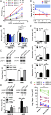
Coligation of LILRB1 is essential for ADE. (A) Binding of LILRB1 to whole DENV or DENV E protein ectodomain. (B) Plaque titers following DENV-2 or ADE infection in the presence of soluble LILRB1 ectodomain (2 µM, 20 µM, 200 µM), 200 µM BSA, or no protein control. (C) Plaque titers following DENV-2 or ADE infection after LILRB1 or FcγRIIB knockdown. Numbers below Western blot indicate levels of proteins relative to LAMP-1. (D) Plaque titers following DENV-2 or ADE infection in THP-1.2R transfected with empty vector or vector expressing LILRB1, mutant LILRB1 (LILRB1MUT), or LILRB4. Numbers below Western blot indicate levels of proteins relative to LAMP-1. (E) Plaque titers following DENV-2–only and ADE infection of primary monocytes treated with sodium stibogluconate (SSG) or PBS control (dashed lines, shaded areas reflect SD). (F) Plaque titers following DENV-1–, -3, or -4–only and ADE infection of primary monocytes treated with SSG (0.138 mM) or PBS control. (G) Plaque titers in primary monocytes derived from PBMCs harvested from 12 healthy individuals and infected in vitro with either DENV-1 (n = 3), DENV-2 (n = 3), DENV-3 (n = 3), or DENV-4 (n = 3) opsonized with h4G2 antibodies at 72 hpi. PBMCs were either pretreated with polyclonal anti-LILRB1 antibody or isotype antibody control. Data are expressed as mean ± SD from three independent experiments. **P < 0.01, *P < 0.05.

Comment in

References

    1. Bhatt S, et al. The global distribution and burden of dengue. Nature. 2013;496(7446):504–507. - PMC - PubMed
    1. Halstead SB, O’Rourke EJ. Dengue viruses and mononuclear phagocytes. I. Infection enhancement by non-neutralizing antibody. J Exp Med. 1977;146(1):201–217. - PMC - PubMed
    1. Simmons CP, et al. Maternal antibody and viral factors in the pathogenesis of dengue virus in infants. J Infect Dis. 2007;196(3):416–424. - PMC - PubMed
    1. Halstead SB. Pathogenesis of dengue: Challenges to molecular biology. Science. 1988;239(4839):476–481. - PubMed
    1. Halstead SB, O’Rourke EJ. Antibody-enhanced dengue virus infection in primate leukocytes. Nature. 1977;265(5596):739–741. - PubMed

Publication types

MeSH terms