. 2014 Mar 14;3(1):R13-30.
doi: 10.1530/EC-13-0103. Print 2014.

Therapeutic management of hypophosphatemic rickets from infancy to adulthood

Affiliations

Therapeutic management of hypophosphatemic rickets from infancy to adulthood

Agnès Linglart et al. Endocr Connect. .

Abstract

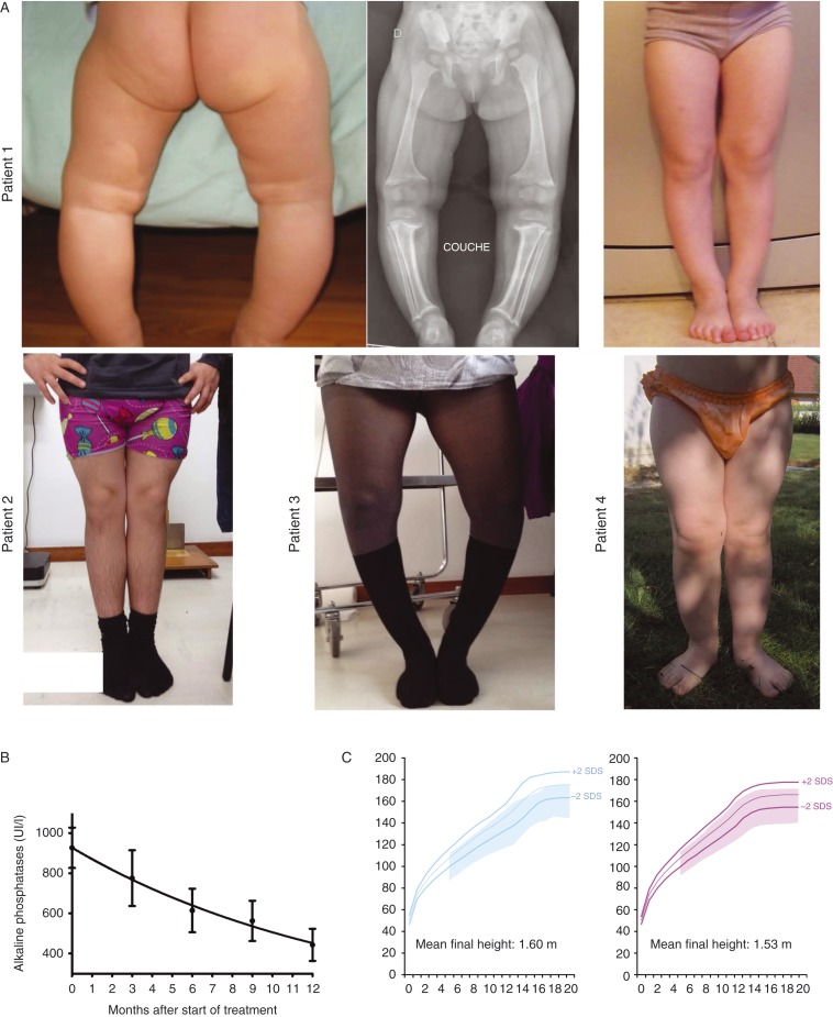
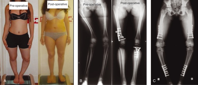
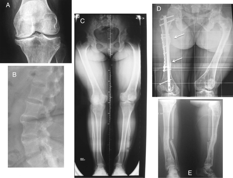
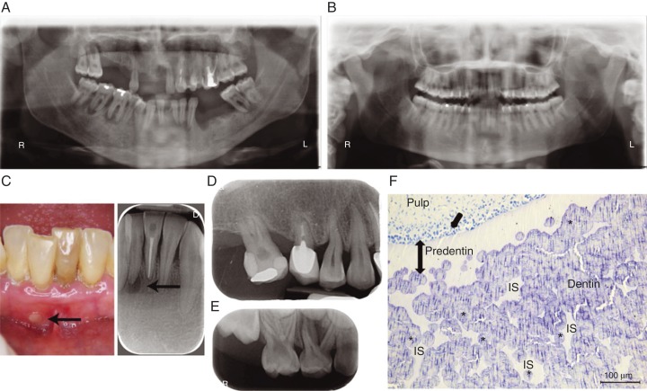
In children, hypophosphatemic rickets (HR) is revealed by delayed walking, waddling gait, leg bowing, enlarged cartilages, bone pain, craniostenosis, spontaneous dental abscesses, and growth failure. If undiagnosed during childhood, patients with hypophosphatemia present with bone and/or joint pain, fractures, mineralization defects such as osteomalacia, entesopathy, severe dental anomalies, hearing loss, and fatigue. Healing rickets is the initial endpoint of treatment in children. Therapy aims at counteracting consequences of FGF23 excess, i.e. oral phosphorus supplementation with multiple daily intakes to compensate for renal phosphate wasting and active vitamin D analogs (alfacalcidol or calcitriol) to counter the 1,25-diOH-vitamin D deficiency. Corrective surgeries for residual leg bowing at the end of growth are occasionally performed. In absence of consensus regarding indications of the treatment in adults, it is generally accepted that medical treatment should be reinitiated (or maintained) in symptomatic patients to reduce pain, which may be due to bone microfractures and/or osteomalacia. In addition to the conventional treatment, optimal care of symptomatic patients requires pharmacological and non-pharmacological management of pain and joint stiffness, through appropriated rehabilitation. Much attention should be given to the dental and periodontal manifestations of HR. Besides vitamin D analogs and phosphate supplements that improve tooth mineralization, rigorous oral hygiene, active endodontic treatment of root abscesses and preventive protection of teeth surfaces are recommended. Current outcomes of this therapy are still not optimal, and therapies targeting the pathophysiology of the disease, i.e. FGF23 excess, are desirable. In this review, medical, dental, surgical, and contributions of various expertises to the treatment of HR are described, with an effort to highlight the importance of coordinated care.

Keywords: X-linked hypophosphatemic rickets; bone; calcium; rare diseases/syndromes.


Figures

Figure 1
Figure 1
Evolution of clinical (leg bowing and growth) and biochemical parameters (alkaline phosphatase levels) during treatment with vitamin D analogs and phosphate supplements in children. (A) Patient 1 is a 2-year-old girl (left an middle panels) recently diagnosed with XLHR and a de novo mutation of PHEX. The same girl is shown at the age of 5 years with straight legs (right panel). Patient 2 is a 14-year-old boy who was treated since the age of four (XLHR and a de novo mutation of PHEX). Patient 3 is a 13-year-old girl who presents with persistent leg bowing despite being treated since she was 3 years old (XLHR and a de novo mutation of PHEX). Patient 4 is a 2-year-old girl who started therapy at the age of 4 months. Diagnosis of XLHR was made in the context of familial disease (mother and two sisters affected). (B) Evolution of alkaline phosphatase levels throughout the first year of therapy in 30 patients affected with HR and elevated FGF23. (C) Growth pattern (range between +2 and −2 SDS is shadowed) in 32 girls and 29 boys affected with HR and elevated FGF23 and receiving vitamin D analogs and phosphate supplements throughout childhood and puberty. Mean, +2, and −2 SDS of French reference growth charts are represented by colored lines.
Figure 2
Figure 2
Surgeries in children and adolescents with XLHR. (A) Bilateral lower limbs mal-alignment including distal right femur valgus and proximal left tibia varus – pre- and post-operative aspects (distal right femur varization and proximal left tibia valgization osteotomies). (B) Pre- and post-operative radiological aspect of the patient displayed in (A). (C) Eight-year-old girl with XLHR and severe pre-operative deformations impeding joints mobility. Recurrence of leg bowing after surgery likely due to compliance issues to phosphate and vitamin D analogs. Bilateral medial proximal tibiae epiphysiodesis inducing varus deformations.
Figure 3
Figure 3
Various burdens of the disease in adults leading to resume therapy with phosphate supplements and vitamin D analogs. (A) Osteoarthritis of the knee in a 28-year-old woman with persistent bone deformities after adolescence. (B) Spinal enthesopathies of a 35-year-old patient with XLHR. (C) Lower limb deformities in a young adult requiring corrective surgery. (D) Dramatic consequences of rickets and osteomalacia in a 30-year-old patient who did not receive vitamin D analogs. Arrows show insufficiency fractures. Bone demineralization and hip osteoarthritis are visible. (E) Delayed healing of fibulae fractures in the same patient following corrective surgeries on both tibias.
Figure 4
Figure 4
Dental defects in patients with X-linked hypophosphatemia (XLH). (A) Orthopantomogram of a 35-year-old XLH patient. Note multiple absent teeth and endodontic lesions. (B) Orthopantomogram of a 30-year-old XLH patient that benefited from vitamin D analogs and phosphate supplements during growth with good compliance. No dental or periodontal defects are evident. (C) Intraoral view and corresponding X-ray of an endodontic infection (arrows) affecting the intact central right lower incisor in a 35 year-old XLH patient. (D) Enlarged pulp chambers, prominent pulp horns, radiolucent hypomineralized dentin and endodontic infection in a 6-year-old XLH patient. (E) Alveolar bone loss in a 45-year-old XLH patient. (F) Toluidine blue-stained section of a third molar germ of a 14-year-old female with XLH showing abnormal dentin mineralization. Numerous nonmineralized interglobular spaces (IS) are observed between unmerged calcospherites in the dentin body (document laboratory EA2496, Dental school University Paris Descartes, France). Asterisks indicate calcospherites, single arrow indicates dentin secreting cells, odontoblasts, and double head arrow indicates extent of predentin.
Figure 5
Figure 5
Scanning electron microscopy views (documents laboratory EA2496, Dental school University Paris Descartes, France) of (A) A deciduous molar of a 6-year-old male patient with XLHR, born from a XLH mother, showing that the major bulk of dentin (arrow) is abnormally mineralized. (B) At higher magnification (white rectangle), the dentin appears extremely porous with multiples unmineralized spaces. (C) Permanent molar of an adult XLH patient hich was not treated during growth. All the dentin bulk (arrow) is abnormally mineralized. (D and E). Permanent third molar of a 15-year old male patient with XLH who was treated during growth with a good compliance to therapy but a late onset. Remaining calcospherites are observed in the outer part of dentin (full arrow) corresponding to infancy, whereas good mineralization (dotted arrow) is seen in the inner part of dentin corresponding to the treatment period. (F) Control permanent molar.

References

    1. Feng JQ, Clinkenbeard EL, Yuan B, White KE, Drezner MK. Osteocyte regulation of phosphate homeostasis and bone mineralization underlies the pathophysiology of the heritable disorders of rickets and osteomalacia. Bone. 2013;54:213–221. doi: 10.1016/j.bone.2013.01.046. - DOI - PMC - PubMed
    1. Martin A, David V, Quarles LD. Regulation and function of the FGF23/klotho endocrine pathways. Physiological Reviews. 2012;92:131–155. doi: 10.1152/physrev.00002.2011. - DOI - PMC - PubMed
    1. Bacchetta J, Sea JL, Chun RF, Lisse TS, Wesseling-Perry K, Gales B, Adams JS, Salusky IB, Hewison M. Fibroblast growth factor 23 inhibits extrarenal synthesis of 1,25-dihydroxyvitamin D in human monocytes. Journal of Bone and Mineral Research. 2013;28:46–55. doi: 10.1002/jbmr.1740. - DOI - PMC - PubMed
    1. Farrow EG, Yu X, Summers LJ, Davis SI, Fleet JC, Allen MR, Robling AG, Stayrook KR, Jideonwo V, Magers MJ, et al. Iron deficiency drives an autosomal dominant hypophosphatemic rickets (ADHR) phenotype in fibroblast growth factor-23 (Fgf23) knock-in mice. PNAS. 2011;108:E1146–E1155. doi: 10.1073/pnas.1110905108. - DOI - PMC - PubMed
    1. Imel EA, Peacock M, Gray AK, Padgett LR, Hui SL, Econs MJ. Iron modifies plasma FGF23 differently in autosomal dominant hypophosphatemic rickets and healthy humans. Journal of Clinical Endocrinology and Metabolism. 2011;96:3541–3549. doi: 10.1210/jc.2011-1239. - DOI - PMC - PubMed

LinkOut - more resources