. 2014 Mar 28;289(13):8865-80.
doi: 10.1074/jbc.M113.506790. Epub 2014 Feb 18.

Characterizing WW domain interactions of tumor suppressor WWOX reveals its association with multiprotein networks

Affiliations

Characterizing WW domain interactions of tumor suppressor WWOX reveals its association with multiprotein networks

Mohammad Abu-Odeh et al. J Biol Chem. .

Abstract

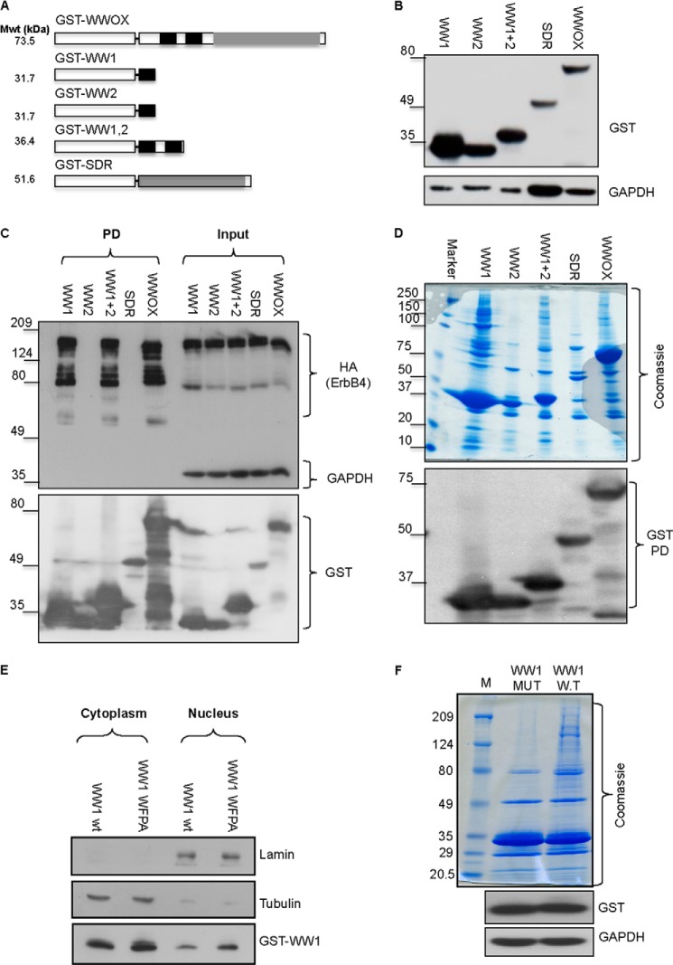
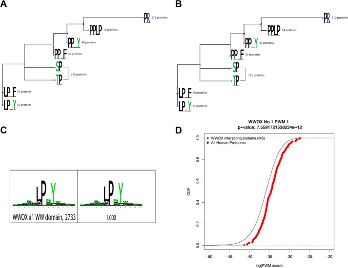
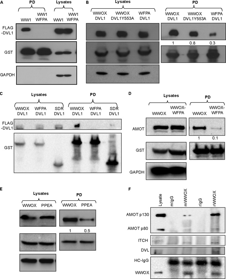
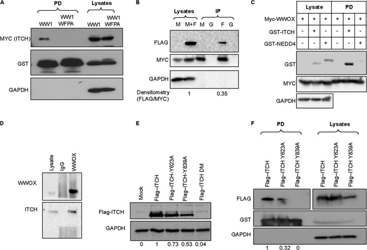
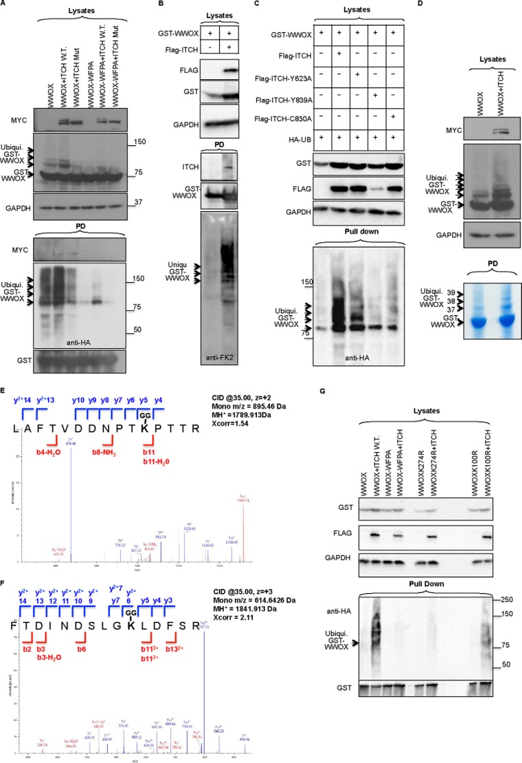
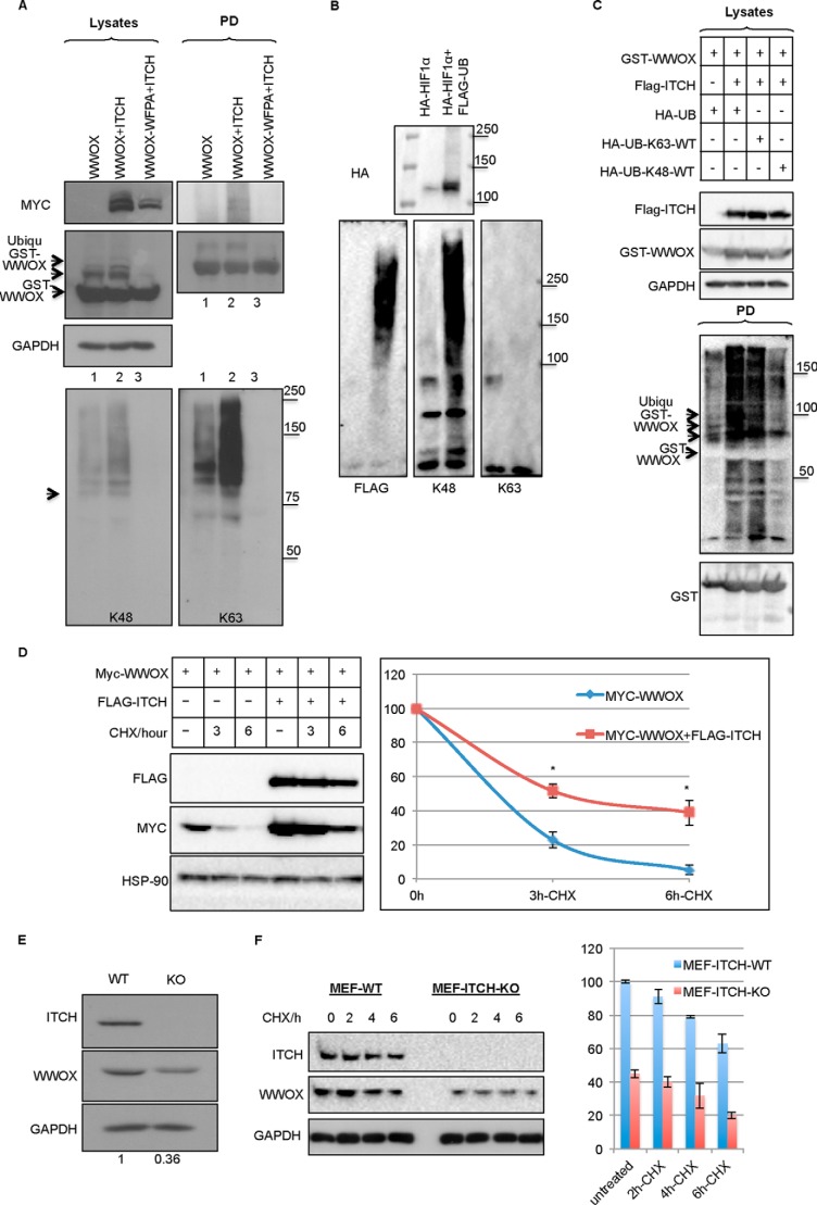
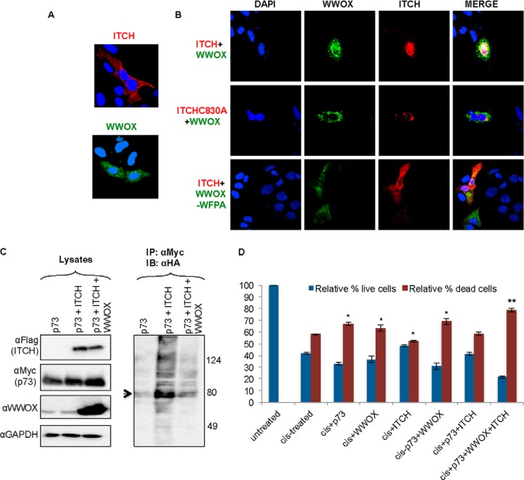
WW domains are small modules present in regulatory and signaling proteins that mediate specific protein-protein interactions. The WW domain-containing oxidoreductase (WWOX) encodes a 46-kDa tumor suppressor that contains two N-terminal WW domains and a central short-chain dehydrogenase/reductase domain. Based on its ligand recognition motifs, the WW domain family is classified into four groups. The largest one, to which WWOX belongs, recognizes ligands with a PPXY motif. To pursue the functional properties of the WW domains of WWOX, we employed mass spectrometry and phage display experiments to identify putative WWOX-interacting partners. Our analysis revealed that the first WW (WW1) domain of WWOX is the main functional interacting domain. Furthermore, our study uncovered well known and new PPXY-WW1-interacting partners and shed light on novel LPXY-WW1-interacting partners of WWOX. Many of these proteins are components of multiprotein complexes involved in molecular processes, including transcription, RNA processing, tight junction, and metabolism. By utilizing GST pull-down and immunoprecipitation assays, we validated that WWOX is a substrate of the E3 ubiquitin ligase ITCH, which contains two LPXY motifs. We found that ITCH mediates Lys-63-linked polyubiquitination of WWOX, leading to its nuclear localization and increased cell death. Our data suggest that the WW1 domain of WWOX provides a versatile platform that links WWOX with individual proteins associated with physiologically important networks.

Keywords: E3 Ubiquitin Ligase; Itch; Mass Spectrometry (MS); Protein-Protein Interactions; Tumor Suppressor Gene; Ubiquitination; WW Domain; WWOX.


Figures

FIGURE 1.
FIGURE 1.
Generation and characterization of WWOX interacting domains. A, schematic illustration of WWOX domains and predicted molecular weight (Mwt). GST is shown as a white box. WW domains of WWOX are shown in black boxes; SDR domains of WWOX are shown as gray boxes. B, generation of full-length and truncated mammalian GST fusions of WWOX. HEK293 cells were transiently transfected with the different GST fusions. After 24 h, cells were lysed, and immunoblot analysis using anti-GST was performed. GAPDH was used for normalization. C, validation of interaction ability of GST-WW1. HEK293 cells were transiently co-transfected with ErbB4-HA and the different GST fusions. At 24 h, cells were lysed, and GST pull-down (PD) was performed. Immunoblot analysis using anti-HA (ErbB4), GST, and GAPDH revealed specific interaction between WW1-containing GST fusions. D, characterization of interaction potential of the different GST-WWOX fusions. HEK293 cells were transiently transfected with the different GST fusions. After 24 h, cells were lysed, and GST pull-down was performed. Top, Coomassie Blue staining of the precipitated proteins of the different fusions. Note that the full-length WWOX lane is most similar to WW1,2 and WW1. Bottom, immunoblot of the different GST fusions using anti-GST antibody. E, characterization of WW1 and WW1-WFPA mutant. HEK293 cells were transiently transfected with GST-WW1 or GST-WW1-WFPA mutant. After 24 h, cells were subfractionated into cytoplasmic and nuclear fractions. Immunoblot analysis, using anti-GST antibody, revealed similar levels and subcellular localization of both proteins. Lamin and tubulin were used to indicate successful nuclear and cytoplasmic subfractionation, respectively. F, characterization of interaction potential of the WW1 domain of WWOX. HEK293 cells were transiently transfected with GST-WW1 or GST-WW1-WFPA mutant. After 24 h, cells were lysed, and GST pull-down was performed. Top, Coomassie Blue analysis of the precipitated proteins of the different fusions. Note that GST-WW1-WFPA (MUT) lost the potential of precipitating protein complexes as compared with intact WW1 (W.T). The lower blots show expression levels of the GST fusions.
FIGURE 2.
FIGURE 2.
A and B, tree motif analysis for putative interacting partners of WWOX obtained from the MS study. The tree was built based on the alignment of eight different motifs of interest. We have used MAFFT to align the sequences and then Geneious to build the tree (neighbor-joining method). The number beside each logo indicates the number of proteins with the motif from our mass spectrometry data (of 240 proteins; MS-SET-1 (A); or of the 144 proteins; MS-SET-2 (B)). C, sequence logo for WW1 domain from phage display. High affinity peptides were identified by phage display, and subsequent sequencing of the phage colonies was performed. These sequences were then filtered, aligned, and clustered using the MUSI software to obtain a sequence logo. D, obtained WW1 binding partners are enriched in hits to the phage-derived specificity profile. Distributions of similarity scores to the phage-derived profile are shown in the observed WW1 binders as compared with the whole proteome. The observed binders are highly enriched in sequences that match well to the phage-derived logo when compared with the background. The p value as from a rank sum test is indicated.
FIGURE 3.
FIGURE 3.
Validation of putative PPXY-containing partners of WWOX identified by MS. A, HEK293 cells were transiently cotransfected with FLAG-DVL1 and GST-WW1 or GST-WW1-WFPA. After 24 h, cells were lysed, and GST pull-down was performed. Precipitates and lysates (input) were immunoblotted with the indicated antibodies. B, HEK293 cells were transiently cotransfected with FLAG-DVL1 or FLAG-DVL1Y553A and GST-WWOX or GST-WWOX-WFPA. After 24 h, cells were lysed, and GST pull-down was performed. Precipitates and lysates (input) were immunoblotted with the indicated antibodies. Numbers below the DVL1 blots represent quantification of bands relative to the GST blot. C, HEK293 cells were transiently cotransfected with FLAG-DVL1 and GST-WWOX or GST-WWOX-WFPA or GST-SDR. Cells were treated as in B. D, HEK293 cells were transiently transfected with FLAG-AMOTL1 and GST-WWOX or GST-WWOX-WFPA. After 24 h, cells were lysed, and GST pull-down was performed. Precipitates and lysates (input) were immunoblotted with the indicated antibodies. Densitometry shows band quantification. E, HEK293 cells were transiently transfected with GST-WWOX and FLAG-AMOTL1 or FLAG-AMOTL1-PPEA. After 24 h, cells were lysed, and GST pull-down was performed. Precipitates and lysates (input) were immunoblotted with the indicated antibodies. Densitometry shows band quantification. F, endogenous interaction. HEK293 cells were lysed and immunoprecipitated using rabbit (r) or mouse (m) anti-WWOX antibody. Precipitates were blotted using anti-WWOX, anti-angiomotin, anti-ITCH, or anti-DVL antibodies. Anti-IgG (r or m) was used as a control.
FIGURE 4.
FIGURE 4.
Physical association between ITCH and WWOX. A, HEK293 cells were transiently cotransfected with MYC-ITCH and GST-WW1 or GST-WW1-WFPA. After 24 h, cells were lysed, and GST pull-down was performed. Precipitates and lysates (input) were immunoblotted with the indicated antibodies. B, HEK293 cells were transiently transfected with MYC-WWOX (M) or MYC-WWOX and FLAG-ITCH (M+F). After 24 h, cells were lysed, and immunoprecipitation (IP) using anti-MYC (M), anti-FLAG (F), or anti-IgG (G) was performed. Precipitates and lysates (input) were immunoblotted with antibodies against FLAG (ITCH) or MYC (WWOX). C, HEK293 cells were co-transfected with MYC-WWOX and GST-ITCH or GST-NEDD4. Cells were treated as in A. Densitometry shows band quantification. D, endogenous interaction. Lysates of HEK293 cells were immunoprecipitated with anti-WWOX or anti-IgG, and complexes were immunoblotted with anti-WWOX and anti-ITCH. E, HEK293 cells were transiently cotransfected with the indicated expression vector (top). At 24 h, cell lysates were probed with anti-FLAG (ITCH). GAPDH was used for normalization. DM, double mutant (Y623A,Y839A). Densitometry shows band quantification. F, HEK293 cells were transiently cotransfected with GST-WWOX and the indicated ITCH expression vector (top). At 24 h, cells were lysed, and GST pull-down was performed. Precipitates and lysates (input) were immunoblotted with the indicated antibodies. Densitometry shows band quantification.
FIGURE 5.
FIGURE 5.
ITCH mediates polyubiquitination of WWOX. A, HEK293 cells were transiently cotransfected with HA-Ub, GST-WWOX or GST-WWOX-WFPA, and MYC-ITCH (WT) or catalytic mutant (Mut) ITCH (C830A). At 24 h, cells were lysed, and GST pull-down was performed. Lysates and precipitates were immunoblotted using the indicated antibodies. The top blots show lysates (input) using the indicated antibodies. GST-WWOX (∼73.5 kDa) and ubiquitinated GST-WWOX protein are observed and indicated by arrows. In the bottom pull-down (PD) blot, arrows indicate ubiquitinated GST-WWOX protein. B, HEK293T cells were transfected with GST-WWOX and FLAG-ITCH as indicated. At 24 h post-transfection, cells were lysed, and GST pull-down was performed overnight. Lysates and pulled down complexes were detected using the indicated antibodies. Anti-FK-2 antibody was used to detect endogenous ubiquitin. C, HEK293 cells were transiently cotransfected with GST-WWOX, HA-Ub, FLAG-ITCH, or FLAG-ITCH-Y623A, FLAG-ITCH-Y839A, or FLAG-ITCH-C830A. At 24 h, cell lysates were subjected to GST pull-down and subsequently probed with anti-HA-HRP. Lysates were immunoblotted with anti-FLAG and anti-GST, and GAPDH was used for normalization. D, HEK293 cells were transiently cotransfected with HA-Ub, MYC-ITCH, and GST-WWOX. At 24 h, cells were lysed, and GST pull-down was performed. The top blots show lysates (input) using the indicated antibodies. GST-WWOX and ubiquitinated GST-WWOX protein are observed. Bottom, Coomassie Blue staining of self-GST pull-down. Bands were digested and submitted to MS analysis. For all panels, an arrow on the left indicates GST-WWOX, whereas arrows on the right indicate ubiquitinated GST-WWOX protein. Numbers indicate sample ID submitted to MS-MS analysis. E and F, annotated MS/MS spectra of the identified WWOX ubiquitinated peptides is shown for LAFTVDDNPTK100PTTR (C) and FTDINDSLGK274LDFSR (D). The Xcorr scores of these peptides are 1.54 and 2.11, and the MH+ masses are 1789.913 and 1841.913, respectively. Matched b and y ions are shown in red and blue, respectively (E). G, HEK293 cells were transiently cotransfected with HA-Ub, MYC-ITCH (WT), GST-WWOX, GST-WWOX-WFPA, GST-WWOX-K274R, or GST-WWOX-K100R. At 24 h, cells were lysed, and GST pull-down was performed. Lysates and precipitates were immunoblotted using the indicated antibodies.
FIGURE 6.
FIGURE 6.
ITCH mediates Lys-63-linked polyubiquitination of WWOX. A, HEK293 cells were transiently cotransfected with HA-Ub, MYC-ITCH, and GST-WWOX or GST-WWOX-WFPA. At 24 h, cells were lysed, and GST pull-down was performed. Lysates and precipitates were immunoblotted using the indicated antibodies. Bottom, immunoblotting with anti-HA (left), anti-Ub-Lys-48 (middle), and anti-Ub-Lys-63 (right). B, HEK293T cells were transfected with HA-HIF1α alone or together with FLAG-Ub. At 24 h post-transfection, cells were treated with 20 μm MG-132 for 4 h. Cells were then lysed, and immunoprecipitation was performed overnight. Lysates and precipitates were detected using anti-FLAG (left), anti-Ub-Lys-48 (middle), and anti-Ub-Lys-63 (right). C, HEK293T cells were transfected with GST-WWOX, FLAG-ITCH, and HA-Ub constructs that are either Lys-63 only or Lys-48 only (all other lysines are mutated to arginines). At 24 h post-transfection, cells were lysed, and GST pull-down was performed overnight. Lysates and pulled down complexes were detected using the indicated antibodies. D and E, ITCH expression is associated with WWOX stabilization. D, HEK293 cells were transiently cotransfected with MYC-WWOX and FLAG-ITCH as indicated at the top. After 24 h, cells were incubated with 20 mg/ml CHX for the indicated times, lysed, and probed with anti-Myc and FLAG antibodies. HSP90 was used for normalization. Quantification of three experiments is shown. p value is 10−5. E, wild-type or Itch-deficient mouse embryonic fibroblasts (MEF) were lysed and immunoblotted with the indicated antibodies. F, wild-type or Itch-deficient mouse embryonic fibroblasts were incubated with 20 mg/ml CHX for the indicated times, lysed, and probed with anti-ITCH and WWOX antibodies. GAPDH was used for normalization. In E and F, densitometry analysis (ratio of WWOX/GAPDH (y axis)) indicates shorter half-life of WWOX in the absence of ITCH. Quantification of three experiments is shown. Data are expressed as mean ± S.E. (error bars), p < 0.001.
FIGURE 7.
FIGURE 7.
WWOX-ITCH functional association. A and B, ITCH coexpression with WWOX mediates WWOX nuclear localization. HeLa cells were transiently transfected with FLAG-ITCH and GFP-WWOX, FLAG-ITCH-C830A and GFP-WWOX, or FLAG-ITCH and GFP-WWOX-WFPA as indicated. At 24 h, cells were fixed and immunostained with anti-FLAG followed by Alexa Fluor-647 secondary conjugated antibodies. Cells were visualized by confocal microscopy at ×60. DAPI, nuclei; red, ITCH; green, GFP-WWOX; yellow, co-localization of WWOX and ITCH. C, WWOX suppresses ITCH-mediated ubiquitination of p73. HEK293 cells were transiently cotransfected with HA-Ub, MYC-p73, FLAG-ITCH, and WWOX as indicated. At 24 h, cells were lysed, and immunoprecipitation (IP) was performed using anti-Myc antibody. Lysates and precipitates were immunoblotted (IB) using the indicated antibodies. Arrow, ubiquitinated p73 (right blot). D, coexpression of WWOX and ITCH enhances p73-mediated cell death. HEK293 cells were transiently transfected with HA-p73, GST-WWOX, or FLAG-ITCH or in the combination indicated. At 24 h, cells were treated with cisplatin (25 μm) for an additional 24 h. The number of live and dead cells was determined using trypan blue. The relative percentage of cells is shown. Shown is the average of three independent experiments. *, p < 0.05; **, p < 0.01. Error bars, S.E.

References

    1. Bork P., Sudol M. (1994) The WW domain: a signalling site in dystrophin? Trends Biochem. Sci. 19, 531–533 - PubMed
    1. Sudol M., Bork P., Einbond A., Kastury K., Druck T., Negrini M., Huebner K., Lehman D. (1995) Characterization of the mammalian YAP (Yes-associated protein) gene and its role in defining a novel protein module, the WW domain. J. Biol. Chem. 270, 14733–14741 - PubMed
    1. Sudol M., Chen H. I., Bougeret C., Einbond A., Bork P. (1995) Characterization of a novel protein-binding module: the WW domain. FEBS Lett. 369, 67–71 - PubMed
    1. Macias M. J., Hyvönen M., Baraldi E., Schultz J., Sudol M., Saraste M., Oschkinat H. (1996) Structure of the WW domain of a kinase-associated protein complexed with a proline-rich peptide. Nature 382, 646–649 - PubMed
    1. Sudol M. (1996) Structure and function of the WW domain. Prog. Biophys. Mol. Biol. 65, 113–132 - PubMed

Publication types