. 2014 Mar 28;289(13):8742-8.
doi: 10.1074/jbc.C113.546713. Epub 2014 Feb 18.

Platelets support extracellular sialylation by supplying the sugar donor substrate

Affiliations

Platelets support extracellular sialylation by supplying the sugar donor substrate

Melissa M Lee et al. J Biol Chem. .

Abstract

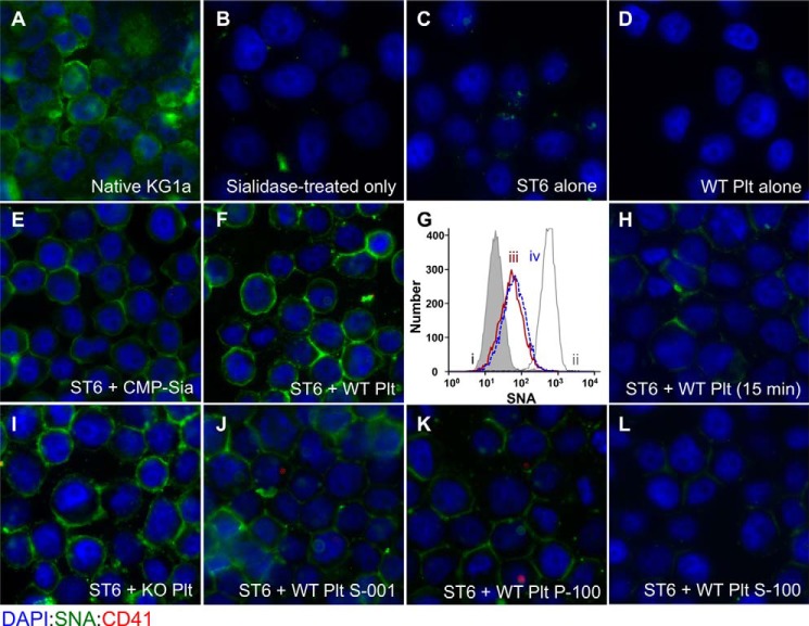
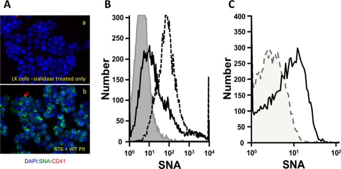
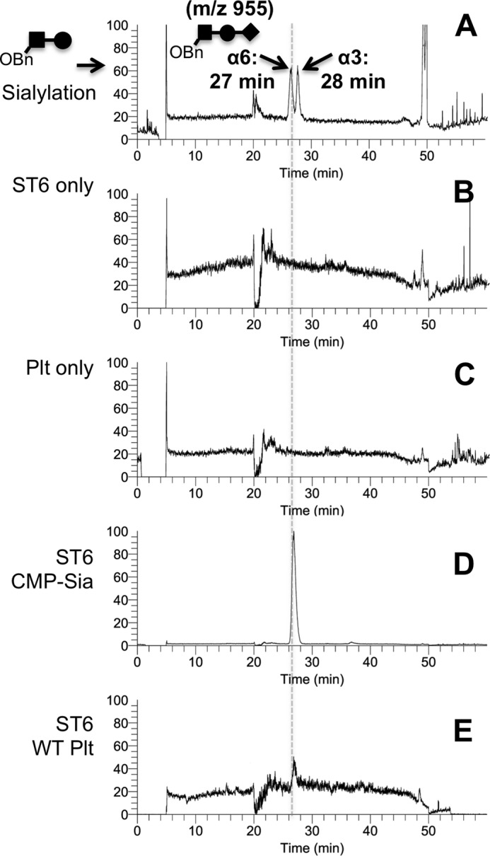
Sizable pools of freely circulating glycosyltransferases are in blood, but understanding their physiologic contributions has been hampered because functional sources of sugar donor substrates needed to drive extracellular glycosylation have not been identified. The blood-borne ST6Gal-1 produced and secreted by the liver is the most noted among the circulatory glycosyltransferases, and decorates marrow hematopoietic progenitor cells with α2,6-linked sialic acids and restricts blood cell production. Platelets, upon activation, secrete a plethora of bioactive molecules including pro- and anti-inflammatory mediators. Cargos of sugar donor substrates for glycosyltransferase activity have also been reported in platelets. Here, we implemented a cell-based system to interrogate platelets for their ability to deliver effectively the sugar donor substrate for extracellular ST6Gal-1 to function. We report that thrombin-activated platelets, at physiologic concentration and pH, can efficiently and effectively substitute for CMP-sialic acid in extracellular ST6Gal-1-mediated sialylation of target cell surfaces. Activated platelets can also supply the sialic acid donor to sialylate the synthetic acceptor, Gal(β1,4)GlcNAcα-o-benzyl, with the product Sia(α2,6)Gal(β1,4)GlcNAcα-o-benzyl structurally confirmed by LC/MS. Platelet-secreted donor substrate was recovered in the 100,000 × g sediment, strongly suggesting the association of this otherwise soluble substrate, putatively CMP-sialic acid, within platelet microparticles. Sequestration within microparticles may facilitate delivery of glycosylation substrate at effective dosages to sites of extracellular glycosylation while minimizing excessive dilution.

Keywords: Cell Surface; Glycosylation; Plasma; Platelets; Serum; Sialyltransferase.


Figures

FIGURE 1.
FIGURE 1.
Activated platelet exudates can substitute for CMP-sialic acid in extrinsic ST6Gal-1-mediated sialylation of target cell surfaces. Fixed native KG1a cells were immobilized onto glass slides, sialidase-treated as described under “Experimental Procedures,” and used as target cells to interrogate extrinsic ST6Gal-1-mediated sialylation of the cell surface. Shown are immunofluorescence images of the target cells after staining with DAPI (blue), SNA (green), and the platelet cell surface marker, CD41 (red). A and B, KG1a cells before and after sialidase treatment, respectively. Unless otherwise specifically noted, sialidase-treated target cells were incubated for 120 min as follows: C, ST6Gal-1 alone; D, wild-type activated platelets (WT-Plt) alone; E, ST6Gal-1 and CMP-sialic acid; and F, ST6Gal-1 and WT-Plt. G shows flow cytometric SNA fluorescence intensities of the target cells that were not immobilized onto slides, as follows: sialidase-treated cells (gray filled in line), sialidase-treated cells upon further treatment with ST6Gal-1 with CMP-Sia (gray line), and ST6Gal-1 with WT- or ST6gal1-KO-Plts (red and blue peaks labeled as ii, iii, and iv, respectively). H, ST6Gal-1 with WT-Plt for only 15 min. I, ST6Gal-1 and St6gal1-KO platelets. J–L, ST6Gal-1 with WT-Plt 1,000 × g supernatant, 100,000 × g pellet, and 100,000 × g supernatant, respectively. All incubations with ST6Gal-1 and platelet fractions, unless stated otherwise specifically, were for 120 min at 37 °C in HEPES buffer at pH 7.4. 225 microunits/ml ST6Gal-1 was used for all except the flow cytometric experiment in G, where 30 microunits/ml was used. CMP-sialic acid was at 100 μm (E). Where platelets or platelet products were used, the equivalent starting amount was 109 platelets/ml except for the flow cytometric experiment (G) where 2.5 × 10⁸ platelets/ml was used.
FIGURE 2.
FIGURE 2.
Cell surface remodeling of primary hematopoietic cells by extrinsic ST6Gal-1 and activated platelets. A, platelet-assisted ST6Gal-1 remodeling of sialidase-treated primary hematopoietic progenitor cells. Hematopoietic progenitors represented by the LK cells from the St6gal1-KO marrow were sialidase-treated, immobilized on glass slides as per the preparation of KG1a cells in Fig. 1, and visualized after staining with DAPI (blue), SNA (green), and the platelet cell surface marker, CD41 (red). Shown are the sialidase-treated LK cells without (panel a) and after incubation with ST6Gal-1 (25 microunits/ml) and thrombin-activated platelets (1 × 10⁸/ml) at 37 °C in RPMI medium for 120 min (panel b). B, extrinsic sialylation of St6gal1-KO LK cells does not require sialidase pretreatment. Native LK cells from the St6gal1-KO marrow were incubated in suspension for 120 min at 37 °C in RPMI medium in the presence of ST6Gal-1 (25 microunits/ml), with 1 × 10⁸/ml thrombin-activated St6gal1-KO platelets (solid line), with 100 μm CMP-sialic acid (dashed line), or with ST6Gal-1 alone (shaded area). C, St6gal1-KO platelets self-sialylate with added ST6Gal-1. 5 × 10⁶ activated St6gal1-KO platelets were incubated for 60 min at 37 °C in RPMI in the presence (solid line) or absence (dashed line) of added ST6Gal-1 (25 microunits/ml).
FIGURE 3.
FIGURE 3.
Activated platelets can replace CMP-sialic acid in the sialylation of Gal(β1,4)GlcNAcα-o-Bn. The conversion of the synthetic acceptor substrate to Sia(α2,6)Gal(β1,4)GlcNAcαo-Bn was monitored by LC/MS, as illustrated using authentic standards in A, where the m/z 955 MS ion that is diagnostic of the trisaccharide yields resolution of α2,6- and α2,3-trisaccharide products on LC, as described under “Experimental Procedures.” B–E, the synthetic acceptor substrate was incubated as follows: B, ST6Gal-1 alone; C, wild-type platelets alone; D, ST6Gal-1 and CMP-sialic acid (5 mm); and E, ST6Gal-1 with wild-type activated platelets (5 × 10⁸/ml). Incubations were performed in 50 mm sodium cacodylate at pH 7.4 for 90 min.

References

    1. Kaplan H. A., Woloski B. M., Hellman M., Jamieson J. C. (1983) Studies on the effect of inflammation on rat liver and serum sialyltransferase: evidence that inflammation causes release of Galβ1→4GlcNAc α2→6 sialyltransferase from liver. J. Biol. Chem. 258, 11505–11509 - PubMed
    1. Dalziel M., Lemaire S., Ewing J., Kobayashi L., Lau J. T. Y. (1999) Hepatic acute phase induction of murine β-galactoside α2,6-sialyltransferase (ST6Gal I) is IL-6 dependent and mediated by elevation of exon H-containing class of transcripts. Glycobiology 9, 1003–1008 - PubMed
    1. Jamieson J. C., Lammers G., Janzen R., Woloski B. M. (1987) The acute phase response to inflammation: the role of monokines in changes in liver glycoproteins and enzymes of glycoprotein metabolism. Comp. Biochem. Physiol. B. 87, 11–15 - PubMed
    1. Bernacki R. J., Kim U. (1977) Concomitant elevations in serum sialytransferase activity and sialic acid content in rats with metastasizing mammary tumors. Science 195, 577–580 - PubMed
    1. Nasirikenari M., Chandrasekaran E. V., Matta K. L., Segal B. H., Bogner P. N., Lugade A. A., Thanavala Y., Lee J. J., Lau J. T. (2010) Altered eosinophil profile in mice with ST6Gal-1 deficiency: an additional role for ST6Gal-1 generated by the P1 promoter in regulating allergic inflammation. J. Leukoc. Biol. 87, 457–466 - PMC - PubMed

Publication types

MeSH terms

Substances