. 2014 Mar 11;111(10):3763-8.
doi: 10.1073/pnas.1320393111. Epub 2014 Feb 18.

Evolutionary history and metabolic insights of ancient mammalian uricases

Affiliations

Evolutionary history and metabolic insights of ancient mammalian uricases

James T Kratzer et al. Proc Natl Acad Sci U S A. .

Abstract

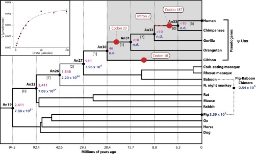
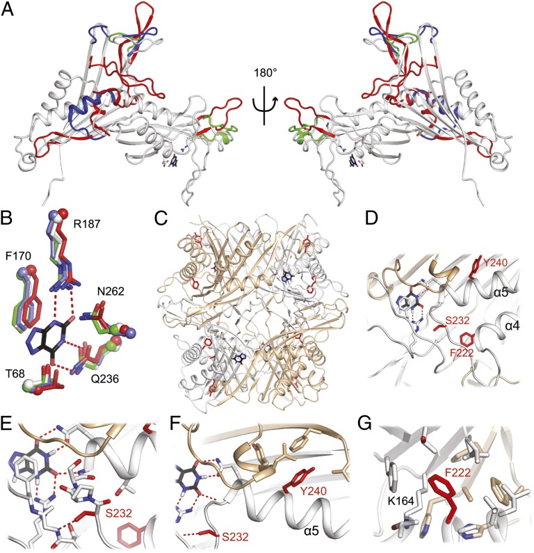
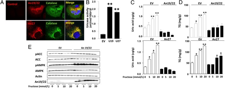
Uricase is an enzyme involved in purine catabolism and is found in all three domains of life. Curiously, uricase is not functional in some organisms despite its role in converting highly insoluble uric acid into 5-hydroxyisourate. Of particular interest is the observation that apes, including humans, cannot oxidize uric acid, and it appears that multiple, independent evolutionary events led to the silencing or pseudogenization of the uricase gene in ancestral apes. Various arguments have been made to suggest why natural selection would allow the accumulation of uric acid despite the physiological consequences of crystallized monosodium urate acutely causing liver/kidney damage or chronically causing gout. We have applied evolutionary models to understand the history of primate uricases by resurrecting ancestral mammalian intermediates before the pseudogenization events of this gene family. Resurrected proteins reveal that ancestral uricases have steadily decreased in activity since the last common ancestor of mammals gave rise to descendent primate lineages. We were also able to determine the 3D distribution of amino acid replacements as they accumulated during evolutionary history by crystallizing a mammalian uricase protein. Further, ancient and modern uricases were stably transfected into HepG2 liver cells to test one hypothesis that uricase pseudogenization allowed ancient frugivorous apes to rapidly convert fructose into fat. Finally, pharmacokinetics of an ancient uricase injected in rodents suggest that our integrated approach provides the foundation for an evolutionarily-engineered enzyme capable of treating gout and preventing tumor lysis syndrome in human patients.

Keywords: evolution; hyperuricemia; pseudogene.


Conflict of interest statement

Conflict of interest statement: E.A.G. declares financial interest because a patent related to this research has been filed by the Georgia Institute of Technology, and this technology is licensed to General Genomics, LLC, which is a start-up founded by E.A.G.. R.J.J. is an inventor on several patents and patent applications related to lowering uric acid as a means for treating hypertension, diabetic nephropathy, and renal disease, and is a founder of XORT, Inc., which is a start-up company that has licensed these applications.

Figures

Fig. 1.
Fig. 1.
Phylogeny of the uricase gene family and enzymatic properties of inferred ancestral uricases. Shown is a phylogenetic tree depicting the evolutionary relationship among mammalian uricase genes. Three genetic lesions are attributed with the pseudogenization of the human uricase gene. A nonsense mutation at codon 33 is common to all great apes (orangutan, gorilla, chimpanzee, and human). An additional nonsense mutation is common for the chimpanzee and human sequences at codon 187. Further, a mutation is located at the splice-acceptor site in intron 2 for the chimpanzee and human sequences. A missense mutation at codon 18 of lesser apes is independent of the great ape uricase pseudogenization event. The ancestral nodes (An) on the tree are labeled in bold. The protein sequences at nodes 19 and 22 are identical and labeled as An19/22. In addition, the protein sequences at nodes 32 and 33 are identical and labeled as An32/33. The numbers in brackets between nodes represent the number of amino acid replacements that occur along each branch. The two stop codons in a full-length human uricase gene were replaced with arginine codons but this recombinant protein exhibited properties analogous to An32/33. Enzymatically characterized uricases are denoted by solid black circles. The specific activity is the amount of uric acid substrate oxidized per minute per milligram of total insoluble protein (shown in purple). Catalytic efficiency was also determined for modern and ancient uricases when possible (shown in blue). (Inset) Example kinetic reaction as monitored by following the oxidation of uric acid. (*kcat/KM for An19/22 is significantly greater than An26’s, P < 0.0001; #kcat/KM for An26 is significantly greater than An27’s, P < 0.0001.) Divergence times for ancestral nodes are provided along the x axis (49).
Fig. 2.
Fig. 2.
Structural features of an ancient mammalian uricase. (A) Superposition of the Anc19/22 structure (gray) on uricases from Bacillus sp. (red; PDB ID code 1J2G), Aspergillus flavus (green; PDB ID code 2YZE), and Arthrobacter globiformis (blue; PDB ID code 1R56). Structurally conserved regions are shown in gray, and divergent regions are highlighted red, green, and blue, respectively. For reference, uric acid from holo A. flavus uricase (PDB ID code 3OBP) is modeled as black sticks; active site residues are shown as white, side-chain sticks. (B) Close-up view of the active site with side chains shown as sticks (N, blue; O, red) and the α-carbon shown as a sphere. (C) Global and close-up view of the tetramer showing the location of the three large-effect amino acid replacements in red. Monomers A and C are colored white, whereas B and D are colored tan. (DG) Close-up views of S232, Y240, and F222, respectively.
Fig. 3.
Fig. 3.
Expression and function of ancestral uricases in human HepG2 cells. (A) Colocalization studies between An19/22 (red, Upper) and An27 (red, Lower) with the peroxisome marker catalase (green). Merged images show colocalization (yellow). Nuclear staining with DAPI (blue). (B) Intracellular uricase activity in control and An19/22- and An27-expressing cells. (C) Intracellular uric acid levels in HepG2 control cells (empty vector; EV) and cells stably expressing An19/22 or An27 and exposed to increasing levels of fructose (from 0 to 20 mM). (D) Intracellular triglyceride levels in HepG2 control cells (EV) and cells stably expressing An19/22 or An27 and exposed to increasing levels of fructose (from 0 to 20 mM). (*P < 0.05 and **P < 0.01 vs. no fructose.) (E) Representative Western blot of lysates from HepG2 cells stably expressing an empty vector control or An19/22. ACC, Acetyl-Coa carboxylase; AMPK, AMP kinase; P, phosphorylated protein.

Comment in

References

    1. Kahn K, Tipton PA. Spectroscopic characterization of intermediates in the urate oxidase reaction. Biochemistry. 1998;37(33):11651–11659. - PubMed
    1. Johnson RJ, et al. Lessons from comparative physiology: Could uric acid represent a physiologic alarm signal gone awry in western society? J Comp Physiol B. 2009;179(1):67–76. - PMC - PubMed
    1. Keebaugh AC, Thomas JW. The evolutionary fate of the genes encoding the purine catabolic enzymes in hominoids, birds, and reptiles. Mol Biol Evol. 2010;27(6):1359–1369. - PMC - PubMed
    1. Hayashi S, Fujiwara S, Noguchi T. Evolution of urate-degrading enzymes in animal peroxisomes. Cell Biochem Biophys. 2000;32(Spring):123–129. - PubMed
    1. Ramazzina I, Folli C, Secchi A, Berni R, Percudani R. Completing the uric acid degradation pathway through phylogenetic comparison of whole genomes. Nat Chem Biol. 2006;2(3):144–148. - PubMed

Publication types

MeSH terms

Associated data