. 2014 Mar 4;111(9):3389-94.
doi: 10.1073/pnas.1321621111. Epub 2014 Feb 18.

Structures of the PutA peripheral membrane flavoenzyme reveal a dynamic substrate-channeling tunnel and the quinone-binding site

Affiliations

Structures of the PutA peripheral membrane flavoenzyme reveal a dynamic substrate-channeling tunnel and the quinone-binding site

Harkewal Singh et al. Proc Natl Acad Sci U S A. .

Abstract

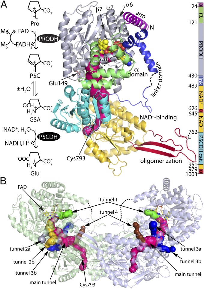
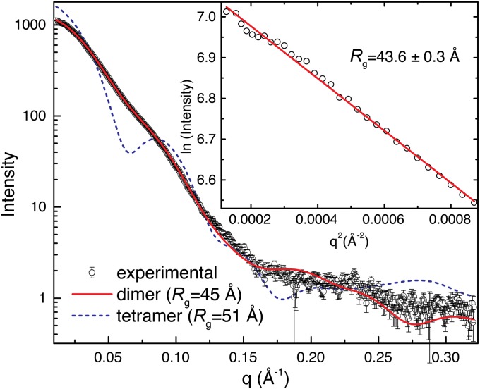
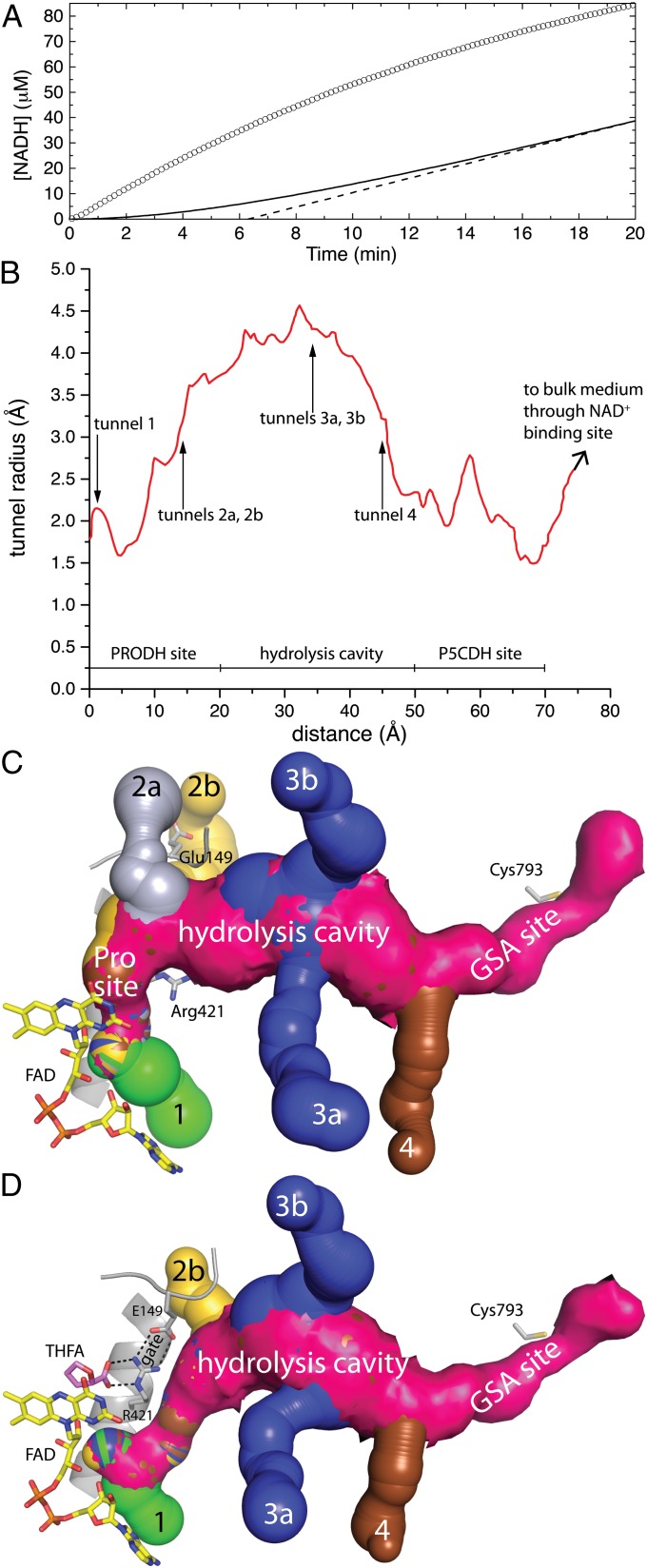
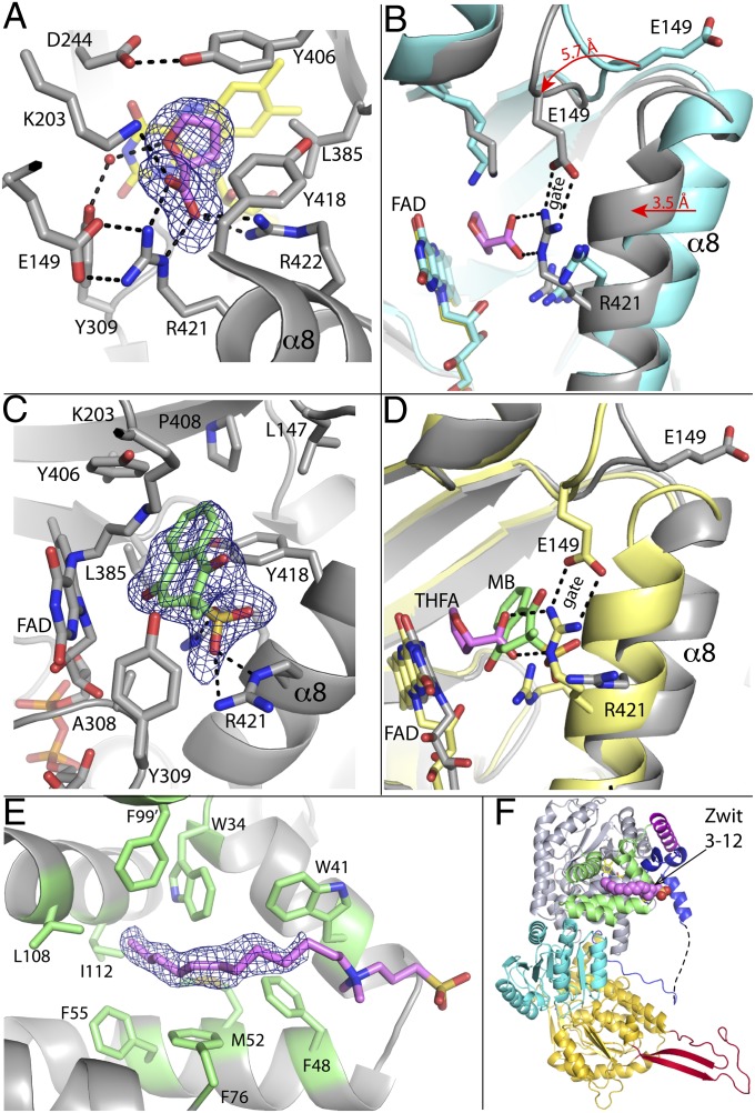
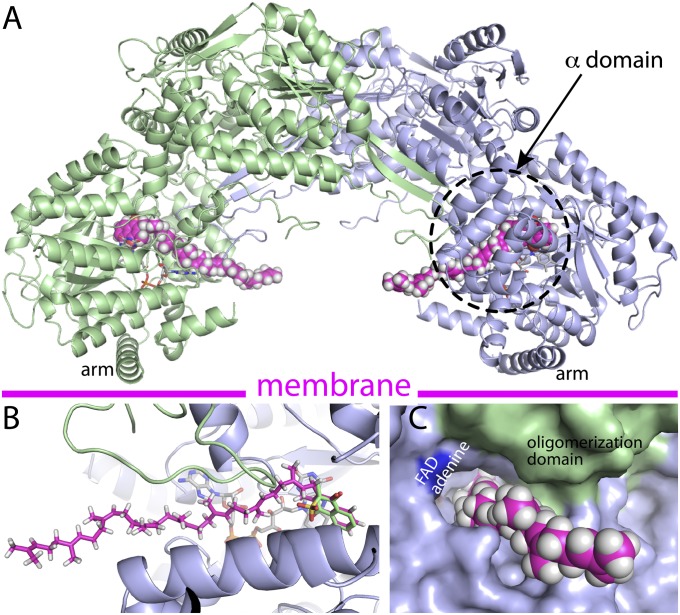
Proline utilization A (PutA) proteins are bifunctional peripheral membrane flavoenzymes that catalyze the oxidation of L-proline to L-glutamate by the sequential activities of proline dehydrogenase and aldehyde dehydrogenase domains. Located at the inner membrane of Gram-negative bacteria, PutAs play a major role in energy metabolism by coupling the oxidation of proline imported from the environment to the reduction of membrane-associated quinones. Here, we report seven crystal structures of the 1,004-residue PutA from Geobacter sulfurreducens, along with determination of the protein oligomeric state by small-angle X-ray scattering and kinetic characterization of substrate channeling and quinone reduction. The structures reveal an elaborate and dynamic tunnel system featuring a 75-Å-long tunnel that links the two active sites and six smaller tunnels that connect the main tunnel to the bulk medium. The locations of these tunnels and their responses to ligand binding and flavin reduction suggest hypotheses about how proline, water, and quinones enter the tunnel system and where L-glutamate exits. Kinetic measurements show that glutamate production from proline occurs without a lag phase, consistent with substrate channeling and implying that the observed tunnel is functionally relevant. Furthermore, the structure of reduced PutA complexed with menadione bisulfite reveals the elusive quinone-binding site. The benzoquinone binds within 4.0 Å of the flavin si face, consistent with direct electron transfer. The location of the quinone site implies that the concave surface of the PutA dimer approaches the membrane. Altogether, these results provide insight into how PutAs couple proline oxidation to quinone reduction.

Keywords: X-ray crystallography; membrane association; proline catabolism.


Conflict of interest statement

The authors declare no conflict of interest.

Figures

Fig. 1.
Fig. 1.
Structure of GsPutA. (A) Reactions catalyzed by PutA and a ribbon drawing of the protomer of the resting enzyme (state I in Fig. S1). The pink surface represents the substrate-channeling tunnel. The domains are colored according to the legend on the right. The dashes denote disordered sections of the α and linker domains. (B) Ribbon drawing of the dimer, with the two protomers colored green and blue. The surfaces represent the substrate-channeling tunnel system. The main substrate-channeling tunnel is colored pink. Smaller tunnels that connect the main tunnel to the bulk medium are colored green (tunnel 1), silver (tunnel 2a), yellow (tunnel 2b), blue (tunnels 3a and 3b), and brown (tunnel 4).
Fig. 2.
Fig. 2.
SAXS analysis of GsPutA. (Inset) A Guinier plot spanning the range of qRg 0.490–1.29; the linear fit has R2 of 0.995. Theoretical curves were calculated from the GsPutA dimer in the asymmetric unit (red) and the BjPutA tetramer (blue dashes).
Fig. 3.
Fig. 3.
Kinetic evidence of substrate channeling and the dynamic tunnel system of GsPutA. (A) Steady-state progress curve of NADH production from proline by GsPutA. The circles represent the experimental curve for the GsPutA PRODH-P5CDH coupled reaction using 0.75 μM GsPutA, 40 mM proline, 150 μM menadione, and 0.2 mM NAD+ (pH 7.5). The solid curve represents the nonchanneling PRODH and P5CDH reaction calculated from a free diffusion model (Eq. S1). The dashed line shows the extrapolation of the nonchanneling model, which yields a transient time of 6.3 min. (B) Plot of the radius of the main tunnel of the resting enzyme as a function of the distance from the flavin calculated using Mole (10). The locations of the ancillary tunnels are indicated. (C) Surface rendering of the tunnel system of the resting enzyme. Note that the Glu149-Arg421 ion pair gate is open. (D) Surface rendering of the tunnel system of the GsPutA–THFA complex. Note that the Glu149-Arg421 ion pair gate is closed, which eliminates tunnel 2a.
Fig. 4.
Fig. 4.
Ligand binding to GsPutA. (A) Electron density and interactions for THFA bound to oxidized GsPutA. The cage represents a simulated annealing σA-weighted FoFc omit map (2.5σ). (B) Comparison of the open (cyan) and THFA-bound closed (gray) PRODH active sites. The arrows show the directions of conformational changes induced by THFA binding. (C) Electron density and interactions for MB bound to NPPG-inactivated GsPutA. The cage represents a simulated annealing σA-weighted FoFc omit map (2.5σ). (D) Comparison of the PRODH active sites of GsPutA-THFA (pink THFA, yellow protein) and inactivated GsPutA-MB (green MB, gray protein), highlighting the proximity of the proline and quinone sites and the structural differences involving α8 and Glu149. (E) Electron density for Zwittergent 3-12. The cage represents a simulated annealing σA-weighted FoFc omit map (2σ). The head group is included in this figure to guide the eye but is omitted in the deposited structure. Phe99 is contributed by the α domain of a symmetry-related molecule. (F) The location of Zwittergent 3-12 (pink spheres) in the protomer, which is oriented and colored as in Fig. 1A.
Fig. 5.
Fig. 5.
Model of GsPutA complexed with a biological quinone (MK-8) based on the GsPutA-MB crystal structure. (A) Model of the dimer with two MK-8 molecules bound. (B) Close-up view of the model showing the superposition of the naphthoquinones of MK-8 (magenta) and MB (green). (C) A view of the isoprenoid chain of MK-8 exiting through tunnel 1.

References

    1. Phang JM, Liu W, Hancock C, Christian KJ. The proline regulatory axis and cancer. Front Oncol. 2012;2:60. - PMC - PubMed
    1. Zarse K, et al. Impaired insulin/IGF1 signaling extends life span by promoting mitochondrial L-proline catabolism to induce a transient ROS signal. Cell Metab. 2012;15(4):451–465. - PMC - PubMed
    1. Lee IR, et al. Reactive oxygen species homeostasis and virulence of the fungal pathogen Cryptococcus neoformans requires an intact proline catabolism pathway. Genetics. 2013;194(2):421–433. - PMC - PubMed
    1. Krishnan N, Doster AR, Duhamel GE, Becker DF. Characterization of a Helicobacter hepaticus putA mutant strain in host colonization and oxidative stress. Infect Immun. 2008;76(7):3037–3044. - PMC - PubMed
    1. Nakajima K, et al. Possible involvement of put A gene in Helicobacter pylori colonization in the stomach and motility. Biomed Res. 2008;29(1):9–18. - PubMed

Publication types

LinkOut - more resources