. 2014 Mar 4;111(9):3454-9.
doi: 10.1073/pnas.1320907111. Epub 2014 Feb 18.

Ion conduction and conformational flexibility of a bacterial voltage-gated sodium channel

Affiliations

Ion conduction and conformational flexibility of a bacterial voltage-gated sodium channel

Céline Boiteux et al. Proc Natl Acad Sci U S A. .

Abstract

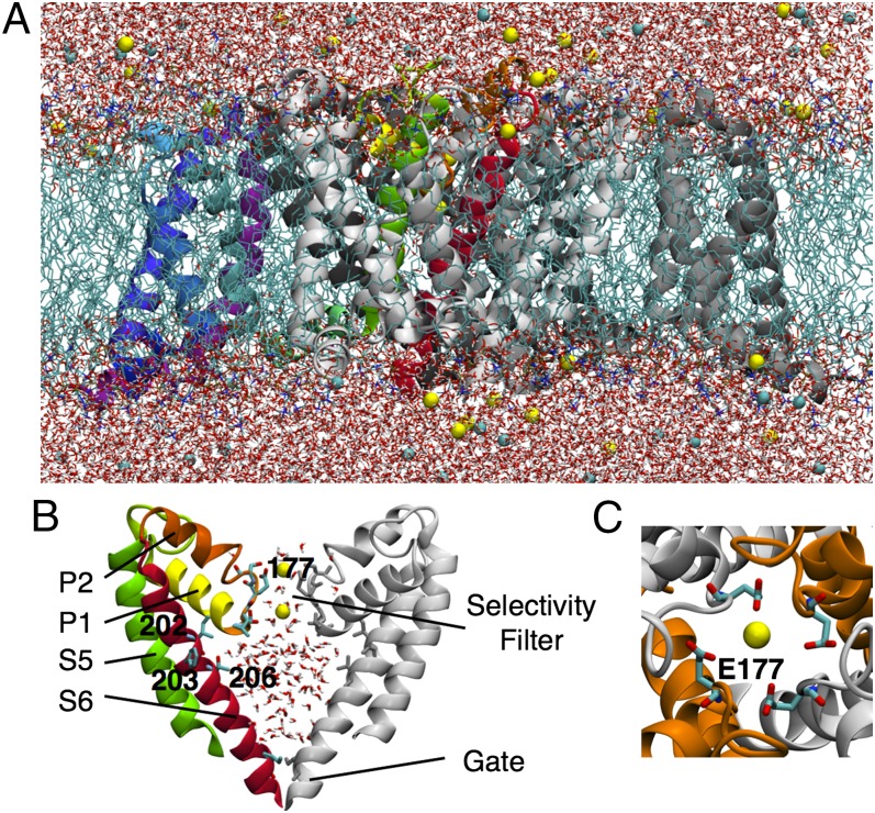
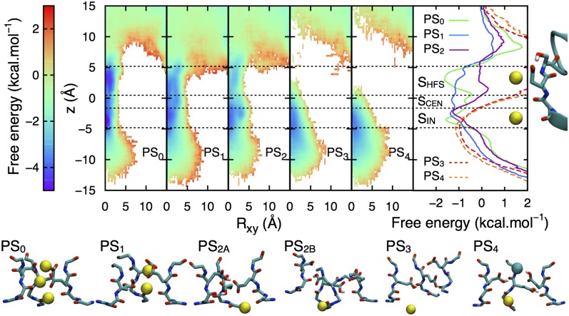
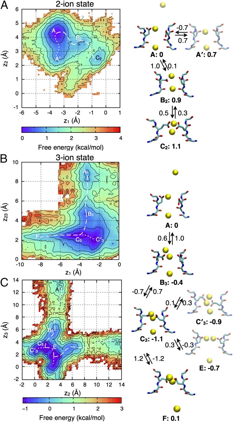
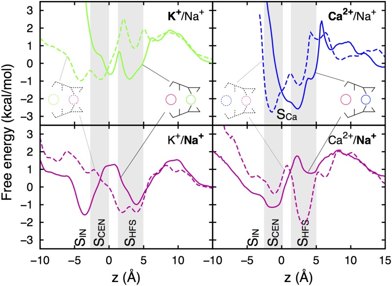
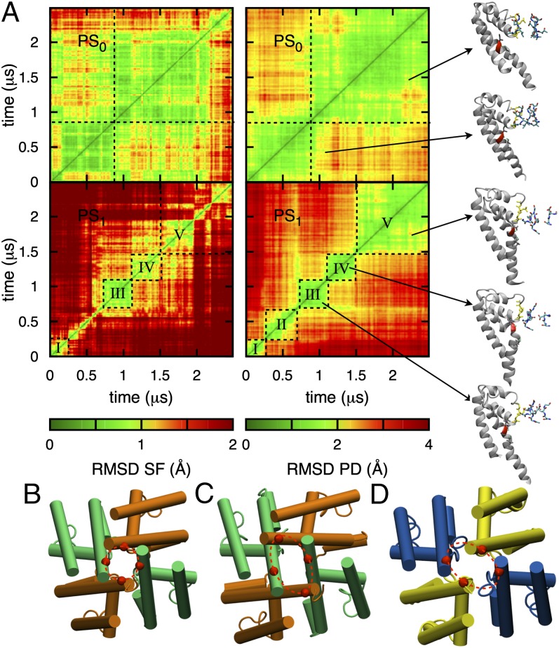
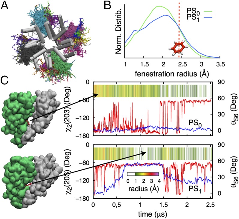
Voltage-gated Na(+) channels play an essential role in electrical signaling in the nervous system and are key pharmacological targets for a range of disorders. The recent solution of X-ray structures for the bacterial channel NavAb has provided an opportunity to study functional mechanisms at the atomic level. This channel's selectivity filter exhibits an EEEE ring sequence, characteristic of mammalian Ca(2+), not Na(+), channels. This raises the fundamentally important question: just what makes a Na(+) channel conduct Na(+) ions? Here we explore ion permeation on multimicrosecond timescales using the purpose-built Anton supercomputer. We isolate the likely protonation states of the EEEE ring and observe a striking flexibility of the filter that demonstrates the necessity for extended simulations to study conduction in this channel. We construct free energy maps to reveal complex multi-ion conduction via knock-on and "pass-by" mechanisms, involving concerted ion and glutamate side chain movements. Simulations in mixed ionic solutions reveal relative energetics for Na(+), K(+), and Ca(2+) within the pore that are consistent with the modest selectivity seen experimentally. We have observed conformational changes in the pore domain leading to asymmetrical collapses of the activation gate, similar to proposed inactivated structures of NavAb, with helix bending involving conserved residues that are critical for slow inactivation. These structural changes are shown to regulate access to fenestrations suggested to be pathways for lipophilic drugs and provide deeper insight into the molecular mechanisms connecting drug activity and slow inactivation.


Conflict of interest statement

The authors declare no conflict of interest.

Figures

Fig. 1.
Fig. 1.
(A) NavAb channel embedded in a hydrated DPPC bilayer (light blue sticks), surrounded by water (red and white sticks) with Na+ and Cl ions (yellow and cyan balls). One of the four monomers shows the VSD in purple/blue and the PD in green/yellow/red. (B) Zoomed view of two monomers of the PD showing S5 (green) and S6 (red), and pore helices P1 (yellow) and P2 (orange) for one of them. The SF and key S6 residues are represented by sticks and labels. (C) Top view of the pore showing the EEEE ring.
Fig. 2.
Fig. 2.
Radial-axial Na+ free energy surfaces for protonation states PS0 to PS4, (with PS2A and PS2B averaged; breakdown provided in SI Appendix, Fig. S3), with 1D projections and initial SF structure shown to the right (binding sites SHFS, SCEN, or SIN indicated with horizontal lines). Dashed curves indicate incomplete sampling for PS3 and PS4. Representative conformations are shown beneath with Na+ and Cl ions as yellow and cyan balls (for clarity only three monomers shown, with all four shown for PS2B to highlight distortion).
Fig. 3.
Fig. 3.
2D PMF projections for Na+ ions in PS0 (see SI Appendix, Fig. S6 for PS1 results), when two (A) or three ions (B and C) occupy the SF, with z23 corresponding to the COM of the upper ions (2, 3). Contouring is at 0.5 kcal/mol, with lowest free energy pathways as dashed curves. The permeation mechanism is shown on the right, with free energies relative to state A and barriers (indicated on the arrows) in kcal/mol (∼±0.5 kcal/mol; negative values implying no barriers).
Fig. 4.
Fig. 4.
PMFs for Na+ (magenta), K+ (green), and Ca2+ (blue) ions during simulations of PS0 (see SI Appendix, Fig. S8 for PS1) in mixtures of Na+/K+ (Left) and Na+/Ca2+ (Right), with two independent simulations shown as solid and dashed curves.
Fig. 5.
Fig. 5.
Fluctuations of the PD. (A) 2D rmsd maps for PS0 (Upper) and PS1 (Lower), considering the SF (backbone of residues 175–179, Left) and PD (backbone of residues 130–220, Right). The black dashed lines delimit blocks of stable conformations (labeled with roman numerals) with representative conformations as insets (SI Appendix, Figs. S5 and S13). Bottom views of the PD in the initial conformation (PDB ID code 3rvy) (B), after 2.5 µs in PS1 (C), and in the proposed inactivated conformation (PDB ID code 4ekw, chains AB) (D), aligned on the S6 helices, are displayed beneath.
Fig. 6.
Fig. 6.
(A) Top view of the PD showing different colored lipids seen within lateral openings during the PS1 simulation. (B) Normalized distributions of fenestration radius for PS0 (green) and PS1 (blue), with red line marking the radius of benzene (∼2.47 Å). (C) Dihedral χ2 of F203 (red curve) and bending angle θS6 (blue curve) for segments D and B in PS0 and PS1, respectively, with corresponding fenestration size as a function of time.

References

    1. Hille B. Ionic Channels of Excitable Membranes. 3rd Ed. Sunderland, MA: Sinauer; 2001.
    1. England S, de Groot MJ. Subtype-selective targeting of voltage-gated sodium channels. Br J Pharmacol. 2009;158(6):1413–1425. - PMC - PubMed
    1. Mike A, Lukacs P. The enigmatic drug binding site for sodium channel inhibitors. Curr Mol Pharmacol. 2010;3(3):129–144. - PubMed
    1. Ren D, et al. A prokaryotic voltage-gated sodium channel. Science. 2001;294(5550):2372–2375. - PubMed
    1. Koishi R, et al. A superfamily of voltage-gated sodium channels in bacteria. J Biol Chem. 2004;279(10):9532–9538. - PubMed

Publication types