. 2014 Feb 13;10(2):e1003922.
doi: 10.1371/journal.ppat.1003922. eCollection 2014 Feb.

The consequences of reconfiguring the ambisense S genome segment of Rift Valley fever virus on viral replication in mammalian and mosquito cells and for genome packaging

Affiliations

The consequences of reconfiguring the ambisense S genome segment of Rift Valley fever virus on viral replication in mammalian and mosquito cells and for genome packaging

Benjamin Brennan et al. PLoS Pathog. .

Abstract

Rift Valley fever virus (RVFV, family Bunyaviridae) is a mosquito-borne pathogen of both livestock and humans, found primarily in Sub-Saharan Africa and the Arabian Peninsula. The viral genome comprises two negative-sense (L and M segments) and one ambisense (S segment) RNAs that encode seven proteins. The S segment encodes the nucleocapsid (N) protein in the negative-sense and a nonstructural (NSs) protein in the positive-sense, though NSs cannot be translated directly from the S segment but rather from a specific subgenomic mRNA. Using reverse genetics we generated a virus, designated rMP12:S-Swap, in which the N protein is expressed from the NSs locus and NSs from the N locus within the genomic S RNA. In cells infected with rMP12:S-Swap NSs is expressed at higher levels with respect to N than in cells infected with the parental rMP12 virus. Despite NSs being the main interferon antagonist and determinant of virulence, growth of rMP12:S-Swap was attenuated in mammalian cells and gave a small plaque phenotype. The increased abundance of the NSs protein did not lead to faster inhibition of host cell protein synthesis or host cell transcription in infected mammalian cells. In cultured mosquito cells, however, infection with rMP12:S-Swap resulted in cell death rather than establishment of persistence as seen with rMP12. Finally, altering the composition of the S segment led to a differential packaging ratio of genomic to antigenomic RNA into rMP12:S-Swap virions. Our results highlight the plasticity of the RVFV genome and provide a useful experimental tool to investigate further the packaging mechanism of the segmented genome.


Conflict of interest statement

The authors have declared that no competing interests exist.

Figures

Figure 1
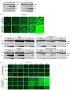
Figure 1. Creation of rMP12:S-Swap.
A. Schematic of the transcription and replication products of the S segments of rMP12 and rMP12:S-Swap. The sites at which oligonucleotides 1 and 2 anneal are indicated. B. Agarose gel showing RT-PCR products to confirm structure of S segment. BHK-21 cells were infected with rMP12 (MP12) or rMP12:S-Swap (SWAP) viruses at an MOI of 1. Total cellular RNA was extracted at 48 h p.i., and S-segment RT-PCR was performed. As a control, PCR on the appropriate cDNA-containing plasmids was performed with the same primers. C. Titres of recombinant virus stocks from multiple independent preparations were determined by plaque assay in BHK-21 cells. The mean titre and standard error of n = 4 preparations of each recombinant virus stock are shown (* p>0.05) D. Comparison of plaque sizes of rMP12, rMP12:S-Swap, rMP12ΔNSs:eGFP or rMP12:S-SwapΔNSs:eGFP on BHK-21 cells. Cell monolayers were fixed at 96 h p.i. with 4% paraformaldehyde and stained with Giemsa solution.
Figure 2
Figure 2. Growth properties of recombinant viruses.
A. Viral growth curves in BHK-21 and A549 cells infected with rMP12 or rMP12:S-Swap (MOI of 0.01 or 5 as indicated). B. The effect of multiplicity of infection on viral yield in BHK-21 cells. Cells were infected with rMP12 or rMP12:S-Swap at multiplicities from 0.0005 to 5 PFU/cell. Viral supernatants were harvested at 72 h p.i. and titrated by plaque assay. Graphs are presented for one representative experiment. C. Viral growth curves in mosquito cells. A. albopictus C6/36 and U4.4, and A. aegypti Ae, cells were infected with rMP12, rMP12:S-Swap, rMP12ΔNSs:eGFP or rMP12:S-SwapΔNSs:eGFP at MOI of 1; BHK-21 cells were similarly infected as a control.
Figure 3
Figure 3. Protein expression by recombinant viruses.
A. Western blot analysis of proteins synthesised in BHK-21 cells infected with rMP12 (M), rMP12:S-Swap (S), rMP12ΔNSs:eGFP (MΔ) or rMP12:S-SwapΔNSs:eGFP (SΔ) at MOI of 1. Cell lysates, prepared at the indicated h p.i., were fractionated by SDS-PAGE, and after transfer, membranes were reacted with rabbit antibodies specific for N, NSs, Gn or eGFP as indicated. Anti-tubulin antibodies were used as a loading control. B. eGFP fluorescence in BHK-21 infected with rMP12ΔNSs:eGFP or rMP12:S-SwapΔNSs:eGFP as above. C. Western blot analysis of infected mosquito cells. A.albopictus C6/36, U4.4 or A. aegypti Ae cells were infected with recombinant viruses (MOI of 1) and lysates prepared at different times post infection. Fractionated proteins were probed with the indicated antibodies. D. eGFP fluorescence in mosquito cells infected with rMP12ΔNSs:eGFP or rMP12:S-SwapΔNSs:eGFP as above. Note that eGFP fluorescence in parts B and D was recorded first and then the same cells were harvested for the western blotting.
Figure 4
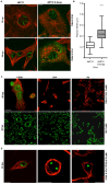
Figure 4. Intracellular localization of NSs in rMP12- or rMP12:S-Swap-infected cells.
A. Vero-E6 cells were infected at MOI of 1, and at the time points indicated the cells were fixed with 4% paraformaldehyde, followed by co-staining with anti-NSs (green) and anti-tubulin (red) antibodies. Cells were examined with a Zeiss LSM confocal microscope. B. Width of NSs filaments. The widths of NSs filaments (50) in rMP12- or rMP12:S-Swap were measured using the LSM Image Browser Software (Carl Zeiss MicroImaging GmbH) and the results presented as the mean width of the filaments and SEM of the two groups (**** = p<0.0001; see Methods). C. Detection of NSs in mosquito cells infected with rMP12. A.albopictus C6/36, U4.4 or A. aegypti Ae cells were infected with recombinant viruses (MOI of 1), and at 48 h p.i. the cells were fixed with 4% paraformaldehyde, followed by co-staining with anti-NSs (green) and anti-tubulin (red) antibodies (upper panels). Duplicate monolayers were stained with anti-N antibodies (lower panels). D. Detection of NSs in mosquito cells infected with rMP12:S-Swap. Cells were infected and stained with anti-NSs (green) and anti-tubulin (red) antibodies as above.
Figure 5
Figure 5. Serial passage of mosquito cells infected with rMP12 or rMP12:S-Swap.
A. albopictus C6/36, U4.4 cells or A. aegypti Ae were infected with rMP12 or rMP12:S-Swap at a MOI of 0.01. Cell monolayers were passaged (split ratio 1∶5) every 5–7 days (when 100% confluency was observed). Cell extracts were prepared from each passage, proteins fractionated SDS-PAGE, transferred to a membrane, and blots probed with anti-N, anti-NSs and anti-tubulin antibodies as indicated. C3/36 cells infected with rMP12:S-Swap died after passage 3.
Figure 6
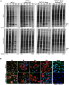
Figure 6. Inhibition of host cell protein and RNA synthesis.
A. Protein synthesis. A549 or A549 NPro cells were infected with rMP12, rMP12:S-Swap, rMP12ΔNSs:eGFP or rMP12:S-SwapΔNSs:eGFP at a MOI of 3. Cells were labelled with 30 µCi [35S] methionine/cysteine for 2 h at the time points indicated, and cell extracts were fractionated by SDS-PAGE. The positions of the viral N and NSs proteins are shown. Total lane intensities were measured by densitometry and compared to the mock-infected sample for each virus time course as indicated. B. RNA synthesis. A549 cells were infected with rMP12 (panels a to d) or rMP12:S-Swap (panels e to h) at a MOI of 3. One hour prior to the time points indicated the uridine analogue 5-ethynyl uridine (EU) was added to the medium and then cells were fixed in 4% formaldehyde. Cells were processed using Click-iT RNA AF488 Imaging Kit (newly synthesised RNA stains green), and then reacted with anti-NSs antibodies and secondary goat anti-rabbit Alexa Fluor 633 antibody (red). As controls, mock-infected cells were left untreated (i) or treated with actinomycin D (Act D) at 5 µg/ml for 1 h prior to 5-EU treatment (j).
Figure 7
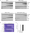
Figure 7. Effect of RVFV NSs on host cell factors.
A. Effect on PKR and p62. A549 cells or A549 cells treated with 5 µg/ml actinomycin D were infected with rMP12 or rMP12:S-Swap at a MOI of 3, or mock-infected. Cell extracts were prepared at the time points indicated, proteins fractionated by SDS-PAGE, and blots probed with anti-N, anti-NSs, anti-PKR, anti-p62, and anti-tubulin antibodies as indicated. B. Induction of interferon. A549 cells were infected with rMP12, rMP12ΔNSs:eGFP, rMP12:S-Swap or rMP12:S-SwapΔNSs:eGFP at MOI of 5, and supernatants harvested at 18 h p.i. After UV treatment, 2-fold dilutions were applied to A549-NPro cells for 24 h, before infection with EMCV. Monolayers were stained with Giesma after a further 96 h. C. The amount of IFN produced is expressed as relative IFN units (RIU), defined as RIU = 2N where N = the number of two-fold dilutions of the supernatants that protected the reporter cells from EMCV challenge.
Figure 8
Figure 8. Analysis of viral RNAs. A. Intracellular RNAs in infected BHK-21 cells.
Cells were infected with rMP12 or rMP12:S-Swap (MOI of 1), and total cell RNA isolated at 48 h p.i. Northern blotting was performed using DIG-labelled probes complementary to the N, NSs, and Gn coding regions in the viral genomic (−) sense RNA and the N coding region in the viral anti-genomic (+) sense RNA. The polarity of the RNA detected by each probe is indicated below the blot: G, genomic sense RNA and AG, anti-genomic sense RNA, defined by the sequence of the 3′/5′ untranslated regions. B. S segment derived mRNAs produced in mosquito cells. A. albopictus C6/36, U4.4 cells or A. aegypti Ae were infected with rMP12 or rMP12:S-Swap at MOI of 1, and total cellular RNA extracted at the indicated times p.i. Northern blotting was performed using the N(+) and NSs(−) probes. C. Analysis of RNA packaged into virions. RNA was extracted purified rMP12 or rMP12:S-Swap grown in BHK-21 cells, and Northern blotting performed with the probes as detailed in (A) above. D. Quantitative RT-PCR analysis of viral RNAs. Total cellular RNA (upper panels) or RNA in purified virus particles (lower panels) of rMP12 or rMP12:S-Swap was analysed using probes specific form the S or M segments as described in Methods. The results are presented as the percentage of genomic RNA species compared to the total RNA of the same segment. Cell line abbreviations: B, BHK-21; C, C6/36; U, U4.4; and A, Ae.

References

    1. Plyusnin A, Beaty BJ, Elliott RM, Goldbach R, Kormelink R, et al. (2012) Bunyaviridae. In: King AMQ, Adams MJ, Carstens EB, Lefkowits EJ, editors. Virus taxonomy: ninth report of the International Committee on Taxonomy of Viruses. London: Elsevier Academic Press. pp. 725–741
    1. Smithburn KC (1949) Rift Valley fever; the neurotropic adaptation of the virus and the experimental use of this modified virus as a vaccine. Br J Exp Pathol 30: 1–16. - PMC - PubMed
    1. Madani TA, Al-Mazrou YY, Al-Jeffri MH, Mishkhas AA, Al-Rabeah AM, et al. (2003) Rift Valley fever epidemic in Saudi Arabia: epidemiological, clinical, and laboratory characteristics. Clin Infect Dis 37: 1084–1092. - PubMed
    1. Gerrard SR, Bird BH, Albariño CG, Nichol ST (2007) The NSm proteins of Rift Valley fever virus are dispensable for maturation, replication and infection. Virology 359: 459–465. - PMC - PubMed
    1. Won S, Ikegami T, Peters CJ, Makino S (2006) NSm and 78-kilodalton proteins of Rift Valley fever virus are nonessential for viral replication in cell culture. J Virol 80: 8274–8278. - PMC - PubMed

Publication types