. 2014 Feb 13;10(2):e1003956.
doi: 10.1371/journal.ppat.1003956. eCollection 2014 Feb.

Male-killing Spiroplasma induces sex-specific cell death via host apoptotic pathway

Affiliations

Male-killing Spiroplasma induces sex-specific cell death via host apoptotic pathway

Toshiyuki Harumoto et al. PLoS Pathog. .

Abstract

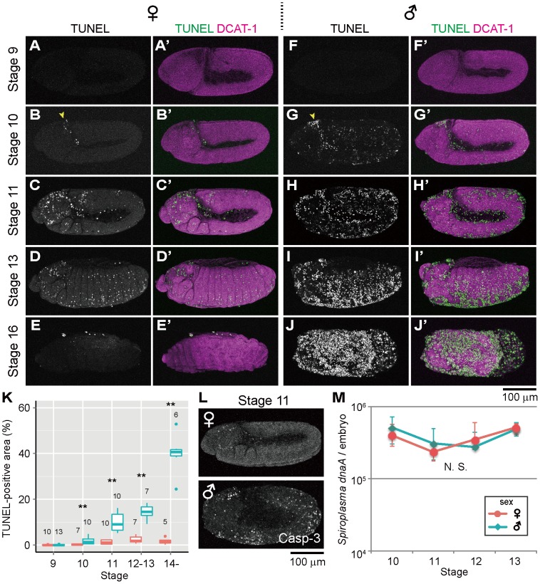
Some symbiotic bacteria cause remarkable reproductive phenotypes like cytoplasmic incompatibility and male-killing in their host insects. Molecular and cellular mechanisms underlying these symbiont-induced reproductive pathologies are of great interest but poorly understood. In this study, Drosophila melanogaster and its native Spiroplasma symbiont strain MSRO were investigated as to how the host's molecular, cellular and morphogenetic pathways are involved in the symbiont-induced male-killing during embryogenesis. TUNEL (terminal deoxynucleotidyl transferase dUTP nick end labeling) staining, anti-cleaved-Caspase-3 antibody staining, and apoptosis-deficient mutant analysis unequivocally demonstrated that the host's apoptotic pathway is involved in Spiroplasma-induced male-specific embryonic cell death. Double-staining with TUNEL and an antibody recognizing epidermal marker showed that embryonic epithelium is the main target of Spiroplasma-induced male-specific apoptosis. Immunostaining with antibodies against markers of differentiated and precursor neural cells visualized severe neural defects specifically in Spiroplasma-infected male embryos as reported in previous studies. However, few TUNEL signals were detected in the degenerate nervous tissues of male embryos, and the Spiroplasma-induced neural defects in male embryos were not suppressed in an apoptosis-deficient host mutant. These results suggest the possibility that the apoptosis-dependent epidermal cell death and the apoptosis-independent neural malformation may represent different mechanisms underlying the Spiroplasma-induced male-killing. Despite the male-specific progressive embryonic abnormality, Spiroplasma titers remained almost constant throughout the observed stages of embryonic development and across male and female embryos. Strikingly, a few Spiroplasma-infected embryos exhibited gynandromorphism, wherein apoptotic cell death was restricted to male cells. These observations suggest that neither quantity nor proliferation of Spiroplasma cells but some Spiroplasma-derived factor(s) may be responsible for the expression of the male-killing phenotype.


Conflict of interest statement

The authors have declared that no competing interests exist.

Figures

Figure 1
Figure 1. Ectopic cell death in Spiroplasma-infected male embryos of D. melanogaster.
(AE) Spiroplasma-infected female embryos and (FJ) Spiroplasma-infected male embryos stained with TUNEL and an antibody against Drosophila α-Catenin (DCAT-1) for visualization of embryonic morphology. Developmental stages are indicated on the left side. Yellow arrowheads indicate the cephalic region at stage 10 where TUNEL positive cells appear first. (K) Quantification of TUNEL-positive area in Spiroplasma-infected female embryos (red) and male embryos (blue). Medians and interquartile ranges are shown with sample sizes (Wilcoxon rank sum test; **, P<0.01). (L) Anti-cleaved Caspase-3 antibody (Casp-3) staining of Spiroplasma-infected female and male embryos at stage 11. (M) Infection dynamics of the male-killing Spiroplasma in developing female embryos (red) and male embryos (blue) from stage 10 to stage 13. Infection densities are indicated in terms of symbiont dnaA gene copies per embryo. Medians and interquartile ranges of 12 measurements are shown. No significant difference was observed between sexes or stages (Kruskal-Wallis test followed by Scheffe test; P>0.05).
Figure 2
Figure 2. Involvement of host's apoptotic pathway in ectopic cell death in Spiroplasma-infected male embryos of D. melanogaster.
(A and B) Quantification of TUNEL-positive area per embryo in control embryos (Ctrl: H99/+ genotype) and apoptosis-deficient embryos (H99: H99/H99 genotype) at stage 11–12 (A) and stage 13–14 (B) (Wilcoxon rank sum test; **, P<0.01). (CJ) TUNEL signals in the Spiroplasma-infected control embryos and apoptosis-deficient embryos at stage 11 (CF) and stage 13 (GJ). The internal edge of the epidermis is highlighted by a dashed line in (E) and (F).
Figure 3
Figure 3. Ectopic cell death in epithelial cells of Spiroplasma-infected male embryos of D. melanogaster.
(A) Schematic diagram of epithelial constitution at early stage 12 when segmentation of embryonic body becomes evident. Each epithelial cell develops a distinct apical-basal polarity. A portion of the apical membrane constitutes the subapical region (SAR; magenta), which shares molecular similarities with the vertebrate tight junction . (BG) Spiroplasma-infected female embryos (BD) and male embryos (EG) at early stage 12 stained with TUNEL and anti-aPKC antibody that visualizes the SAR of epithelial cells. The boxed regions in (B) and (E) are magnified in (C) and (F), respectively. Z-sections of (B) and (E) are shown in (D) and (G), respectively. Intersegmental furrows are indicated by yellow brackets.
Figure 4
Figure 4. Ectopic cell death and neural defects in Spiroplasma-infected male embryos of D. melanogaster.
(A) Schematic diagram of the embryonic nervous system at late stage 12, wherein the central nervous system (CNS) and the peripheral nervous system (PNS) are depicted in red and blue, respectively. (B) Schematic diagram of neurogenesis at the cellular level: blue, neuroblast (or precursor neural cell); green, ganglion mother cells (or GMC); and red, neurons (or differentiated neural cells). (CF) Lateral views of Spiroplasma-infected female embryos (CD) and male embryos (EF) at late stage 12 stained with TUNEL and anti-Elav antibody that visualizes differentiated neural cells. Single optical-sections through CNS of (C) and (E) are shown in (D) and (F), respectively. Green brackets and yellow brackets indicate CNS and PNS, respectively. (GH) Dorsal views of Spiroplasma-infected female embryos (G) and male embryos (H) at stage 10 stained with TUNEL and anti-Krüppel (Kr) antibody that visualizes precursor neural cells or neuroblasts. Single optical sections through the neuroblast layer are selected.
Figure 5
Figure 5. Neural defects in Spiroplasma-infected male embryos of apoptosis-deficient mutant of D. melanogaster.
Spiroplasma-infected female embryos (A and C) and male embryos (B and D) of the apoptosis-deficient H99/H99 genotype at late stage 12 stained with anti-Elav antibody to visualize CNS and PNS. Single optical-sections through CNS of (A) and (B) are shown in (C) and (D), respectively.
Figure 6
Figure 6. Spiroplasma-infected gynandromorphic embryo of D. melanogaster wherein apoptotic cell death is associated with male cells.
This is most likely a stage 13 embryo, although its precise developmental stage cannot be determined because of severe morphological defects. (AC) Images of immunostaining with anti-Sxl antibody by which female cells are specifically stained. (A′C′) Images of immunostaining with anti-cleaved-Casp-3 antibody by which apoptotic cells are selectively visualized. (A″C″) Merged images. The boxed region in (A″) is magnified in (B)–(B″). Z-sections of dotted yellow line in (B″) is shown in (C)–(C″). In (B′), the area of Sxl-expressing female cells is highlighted by dotted yellow line.

References

    1. Buchner P (1965) Endosymbiosis of Animals with Plant Microorganisms. New York, NY, USA: Interscience Publishers. 909 p.
    1. Bourtzis K, Miller TA (2003) Insect Symbiosis. Boca Raton, FL: CRC Press. 156 p.
    1. Moran NA, McCutcheon JP, Nakabachi A (2008) Genomics and evolution of heritable bacterial symbionts. Annu Rev Genet 42: 165–190 doi:10.1146/annurev.genet.41.110306.130119 - DOI - PubMed
    1. Ohkuma M (2003) Termite symbiotic systems: efficient bio-recycling of lignocellulose. Appl Microbiol Biotechnol 61: 1–9 doi:10.1007/s00253-002-1189-z - DOI - PubMed
    1. Oliver KM, Degnan PH, Burke GR, Moran NA (2010) Facultative symbionts in aphids and the horizontal transfer of ecologically important traits. Annu Rev Entomol 55: 247–266 doi:10.1146/annurev-ento-112408-085305 - DOI - PubMed

Publication types