Review
. 2014 Feb 4:5:26.
doi: 10.3389/fphys.2014.00026. eCollection 2014.

The emerging roles of ribosome biogenesis in craniofacial development

Affiliations
Review

The emerging roles of ribosome biogenesis in craniofacial development

Adam P Ross et al. Front Physiol. .

Abstract

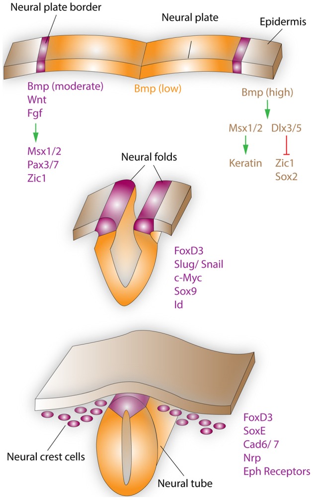
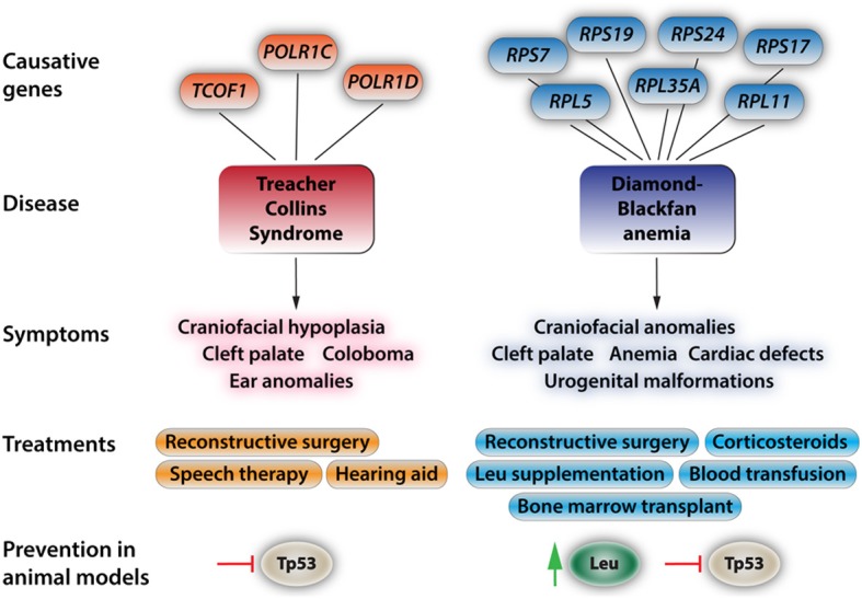
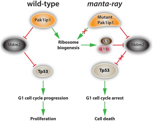
Neural crest cells (NCCs) are a transient, migratory cell population, which originates during neurulation at the neural folds and contributes to the majority of tissues, including the mesenchymal structures of the craniofacial skeleton. The deregulation of the complex developmental processes that guide migration, proliferation, and differentiation of NCCs may result in a wide range of pathological conditions grouped together as neurocristopathies. Recently, due to their multipotent properties neural crest stem cells have received considerable attention as a possible source for stem cell based regenerative therapies. This exciting prospect underlines the need to further explore the developmental programs that guide NCC differentiation. This review explores the particular importance of ribosome biogenesis defects in this context since a specific interface between ribosomopathies and neurocristopathies exists as evidenced by disorders such as Treacher-Collins-Franceschetti syndrome (TCS) and Diamond-Blackfan anemia (DBA).

Keywords: TP53; cell cycle regulation; craniofacial development; neural crest; neurocristopathies; ribosome biogenesis; ribosomopathies.


Figures

Figure 1
Figure 1
Neural crest formation and migration during development. Neural crest regionalization (top) at the boundary of the neural plate and epidermis is a multi-step process. First, the border of the neural plate is set via secretion of neural plate inductive signals (Fgf, Bmp, and Wnt) from the ventral ectoderm and paraxial mesoderm (not shown). Anteriorly, the timing of Bmp and Wnt signaling contributes toward setting the boundaries between epidermis, prospective neural crest, and neural plate. In the narrow band, where Wnt signaling induces Bmp signaling and Wnt signaling is not subsequently turned off, NCCs are formed. Bmp, Wnt, and Fgf, which are secreted by the prospective neural crest, induce the expression of border regionalization genes such as Msx1/2, Pax3/7, and Zic1. In contrast, in the epidermis high concentrations of Bmp induce the expression Msx1/2, which promote keratin expression and Dlx3/5, which induce Zic1 and Sox2 expression. Neural crest specification (middle) starts with the expression of FoxD3, Slug/Snail, c-Myc, Sox9, and Id by the border cells, which prevents this region from becoming either neural plate or epidermal tissue. EMT, delamination, and migration of NCCs (bottom), is primarily induced by FoxD3, Snail, and Sox9. These factors are also capable of inducing a cranial neural crest fate for cells of the lateral neural tube, when ectopically expressed in this region. After delamination NCCs migrate to their respective destinations, regulated by the expression of proteins such as FoxD3, SoxE, Cad6/7, Nrp, and Eph receptors. Specifically, the head and facial structures are largely products of the cranial neural crest, which is a mixed population of cells, with about 10% of these cells being multipotent progenitor cells.
Figure 2
Figure 2
Aspects of the two most prevalent and best-understood neurocristopathies/ribosomopathies, Treacher Collins Syndrome and Diamond-Blackfan anemia. At the top, the diagram depicts known genes with causative mutations in the respective disorders. For more in depth reviews see Trainor (2010), Dauwerse et al. (2011), Payne et al. (2012), and Boultwood et al. (2013). The most-common symptoms are listed below. These symptoms may appear isolated or more commonly in conjunction with the other symptoms. Current treatments and approaches for ameliorating the effects of these syndromes. For the commonly found craniofacial dysplasias only reconstructive surgery is effectively used at this point. While leucine treatment has had positive results in human patients, amino acid treatments come with a bevy of side effects such as ketoacidosis, decreased blood sugar due to increased insulin release by the pancreas, complications of lung function, and hepatic encephalopathy. Recent studies in animal models have shown that Tp53 inhibition will at least partially rescue phenotypic deficiencies in TCS and that Tp53 inhibition and leucine treatment reduce the severity of the effects of DBA.
Figure 3
Figure 3
Proposed mechanism of action of Pak1ip1 in both wild-type and homozygous manta-ray mutants. In the wild-type (left) Pak1ip1 acts as an inhibitor of the E3 ubiquitin ligase Mdm2, which acts as an inhibitor of Tp53 activity through an auto-regulatory feedback loop. Mdm2 targets Tp53 for degradation by the proteasome, blocks its transcriptional activity, and facilitates its export from the nucleus. Low levels of Tp53 activity promote cell cycle progression at the G1 checkpoint and cellular proliferation. In the manta-ray mutant (right) Pak1ip1 causes altered 60S ribosome biogenesis, resulting in nucleolar stress and the accumulation of freely circulating ribosomal proteins L5 and L11, which inhibit Mdm2 activity. This series of events leads to an increase in Tp53 levels, G1 cell cycle arrest, and subsequent cell death, which appears to particularly affect aspects of the cranial neural crest.

References

    1. Abu-Issa R., Smyth G., Smoak I., Yamamura K., Meyers E. N. (2002). Fgf8 is required for pharyngeal arch and cardiovascular development in the mouse. Development 129, 4613–4625 - PubMed
    1. Attie T., Till M., Pelet A., Amiel J., Edery P., Boutrand L., et al. (1995). Mutation of the endothelin-receptor B gene in Waardenburg-Hirschsprung disease. Hum. Mol. Genet. 4, 2407–2409 10.1093/hmg/4.12.2407 - DOI - PubMed
    1. Bastidas F., De Calisto J., Mayor R. (2004). Identification of neural crest competence territory: role of Wnt signaling. Dev. Dyn. 229, 109–117 10.1002/dvdy.10486 - DOI - PubMed
    1. Bohnsack B. L., Lai L., Dolle P., Hirschi K. K. (2004). Signaling hierarchy downstream of retinoic acid that independently regulates vascular remodeling and endothelial cell proliferation. Genes Dev. 18, 1345–1358 10.1101/gad.1184904 - DOI - PMC - PubMed
    1. Bolande R. P. (1997). Neurocristopathy: its growth and development in 20 years. Pediatr. Pathol. Lab. Med. 17, 1–25 10.1080/107710497174994 - DOI - PubMed