

Comparison of mechanical property and role between enamel and dentin in the human teeth
- PMID: 24550998
- PMCID: PMC3924884
- DOI: 10.1177/1758736014520809
Comparison of mechanical property and role between enamel and dentin in the human teeth
Abstract
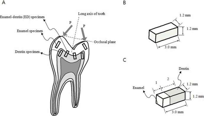
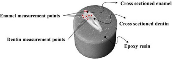
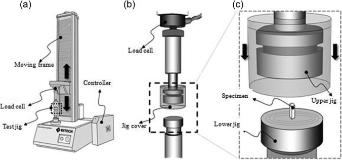
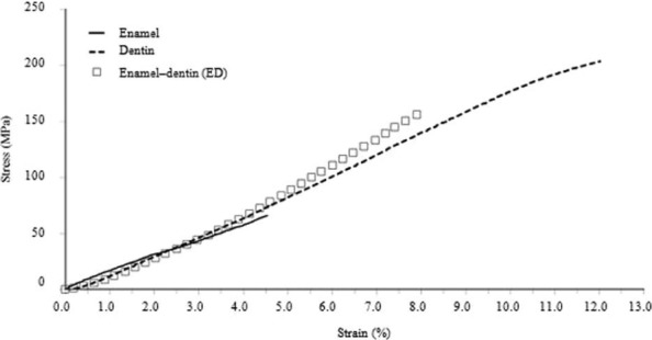
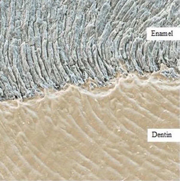
The mechanical properties of enamel and dentin were studied using test specimens having the same shape and dimensions because these properties might vary with the experimental conditions and specimen shapes and dimensions. Healthy human teeth were used as specimens for mechanical tests. The stress (MPa), strain (%), and elastic modulus (E, MPa) of the specimens were obtained from compression tests. The maximum stresses of the enamel, dentin, and enamel-dentin specimens were 62.2 ± 23.8, 193.7 ± 30.6, and 126.1 ± 54.6 MPa, respectively. The maximum strains of the enamel, dentin, and enamel-dentin specimens were 4.5 ± 0.8%, 11.9 ± 0.1%, and 8.7 ± 2.7%, respectively. The elastic moduli of the enamel, dentin, and enamel-dentin specimens were 1338.2 ± 307.9, 1653.7 ± 277.9, and 1628.6 ± 482.7 MPa, respectively. The measured hardness value of enamel specimens (HV = 274.8 ± 18.1) was around 4.2 times higher than that of dentin specimens (HV = 65.6 ± 3.9). Judging from the measured values of the stress and strain of enamel specimens, enamel tended to fracture earlier than dentin; therefore, it was considered more brittle than dentin. However, judging from the measured hardness values, enamel was considered harder than dentin. Therefore, enamel has higher wear resistance, making it suitable for grinding and crushing foods, and dentin has higher force resistance, making it suitable for absorbing bite forces. The different mechanical roles of enamel and dentin may arise from their different compositions and internal structures, as revealed through scanning electron micrographs of enamel and dentin.
Keywords: Mechanical properties; compression tests; dentin; enamel; hardness tests.
Conflict of interest statement
Figures
References
-
- Staines M, Robinson WH, Hood JAA. Spherical indentation of tooth enamel. J Mater Sci 1981; 16(9): 2551–2556
-
- Tillberg A, Järvholm B, Berglund A. Risks with dental materials. Dent Mater 2008; 24(7): 940–943 - PubMed
-
- Vaderhobli RM. Advances in dental materials. Dent Clin North Am 2011; 55(3): 619–625 - PubMed
-
- Cuy JL, Mann AB, Livi KJ, et al. Nanoindentation mapping of the mechanical properties of human molar tooth enamel. Arch Oral Biol 2002; 47: 281–291 - PubMed
-
- Gašperšič D. Micromorphometric analysis of cervical enamel structure of human upper third molars. Arch Oral Biol 1995; 40(5): 453–457 - PubMed
LinkOut - more resources
Full Text Sources
Other Literature Sources