

Role of screening of whole spine with sagittal MRI with MR myelography in early detection and management of occult intrasacral meningocele
- PMID: 24551000
- PMCID: PMC3912767
- DOI: 10.4103/1793-5482.125660
Role of screening of whole spine with sagittal MRI with MR myelography in early detection and management of occult intrasacral meningocele
Abstract
Objective: We evaluated the role of screening of the whole spine by sagittal magnetic resonance imaging (MRI) along with MR myelography in early detection and management of occult intrasacral meningocele.
Materials and methods: A prospective and retrospective analysis of MRI and MR myelography studies of the whole spine over a period of one year was performed.
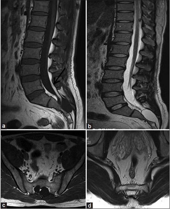
Results: Thirty cases with sacral meningeal cysts were seen. On MRI, six patients (three males, three females) fulfilled the criterion of occult intrasacral meningocele. These patients showed a cyst of cerebrospinal fluid (CSF) signal intensity in the sacral canal below the dural sac. This cyst communicated with the thecal sac through a narrow pedicle. Fat signal intensity in the filum terminale and occult sacral dysraphism in the form of an absent or hypoplastic neural arch was observed in all the patients. Low-lying conus medullaris with thick filum terminale was seen in five of these six patients. Excision of the cyst with the release of filum was performed in two patients with a favorable outcome.
Conclusion: Screening MRI with MR myelography of the whole spine may play a role in the early detection and management of occult intrasacral meningocele. The commonly associated thick filum terminale and low-lying conus medullaris may be missed otherwise that may lead to a progression of symptoms.
Keywords: MR; myelography; occult meningocele.
Conflict of interest statement
Figures


Similar articles
-
Occult intrasacral meningocele with tethered cord--case report.Neurol Med Chir (Tokyo). 1995 May;35(5):321-4. doi: 10.2176/nmc.35.321. Neurol Med Chir (Tokyo). 1995. PMID: 7623956
-
Intrasacral meningocele in the pediatric population.J Neurosurg Pediatr. 2013 Jun;11(6):615-22. doi: 10.3171/2013.3.PEDS12519. Epub 2013 Apr 19. J Neurosurg Pediatr. 2013. PMID: 23601014
-
Meningocele Manqué Discovered During Filum Terminale Resection in Occult Tethered Cord Syndrome.World Neurosurg. 2019 Dec;132:148-153. doi: 10.1016/j.wneu.2019.08.016. Epub 2019 Aug 17. World Neurosurg. 2019. PMID: 31430540
-
Occult, bilateral anterior sacral and intrasacral meningeal and perineurial cysts: case report and review of the literature.Neurosurgery. 1990 Dec;27(6):981-6. doi: 10.1097/00006123-199012000-00020. Neurosurgery. 1990. PMID: 2274142 Review.
-
Terminal syringohydromyelia and occult spinal dysraphism.J Neurosurg. 1994 Oct;81(4):513-9. doi: 10.3171/jns.1994.81.4.0513. J Neurosurg. 1994. PMID: 7931583 Review.
Cited by
-
Magnetic Resonance Imaging in Paediatric Spinal Dysraphism with Comparative Usefulness of Various Magnetic Resonance Sequences.J Clin Diagn Res. 2017 Aug;11(8):TC17-TC22. doi: 10.7860/JCDR/2017/30134.10393. Epub 2017 Aug 1. J Clin Diagn Res. 2017. PMID: 28969239 Free PMC article.
-
[Spinal cysts : Diagnostic workup and therapy].Radiologe. 2018 Feb;58(2):113-119. doi: 10.1007/s00117-017-0350-8. Radiologe. 2018. PMID: 29411052 Review. German.
References
-
- Nabors MW, Pait TG, Byrd EB, Karim NO, Davis DO, Kobrine AI, et al. Updated assessment and current classification of spinal meningeal cysts. J Neurosurg. 1988;68:366–77. - PubMed
-
- Hamamcioglu MK, Hicdonmez T, Kilincer C, Cobanoglu S. Intrasacral extradural arachnoid cysts. Neurol Med Chir (Tokyo) 2008;48:223–6. - PubMed
-
- Choi JY, Kim SH, Lee WS, Sung KH. Spinal extradural arachnoid cyst. Acta Neurochir (Wien) 2006;148:579–85. - PubMed
-
- Doty JR, Thomson J, Simonds G, Rengachary SS, Gunby EN. Occult intrasacral meningocele: Clinical and radiographic diagnosis. Neurosurgery. 1989;24:616–25. - PubMed
-
- Doi H, Toyoda I, Matsumoto K, Imai S, Sekimizu M. Occult intrasacral meningocele with tethered cord - case report. Neurol Med Chir (Tokyo) 1995;35:321–4. - PubMed
LinkOut - more resources
Full Text Sources
Other Literature Sources