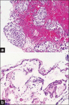
Symptomatic Rathke's cleft cyst with a co-existing pituitary tumor; Brief review of the literature
- PMID: 24551002
- PMCID: PMC3912769
- DOI: 10.4103/1793-5482.125662
Symptomatic Rathke's cleft cyst with a co-existing pituitary tumor; Brief review of the literature
Abstract
Pituitary adenomas and Rathke's cleft cysts (RCCs) share a common embryological origin. Occasionally, these two lesions can present within the same patient. We present a case of a 39-year-old male who was found to have a large sellar lesion after complaints of persistent headaches and horizontal nystagmus. Surgical resection revealed components of a RCC co-existing with a pituitary adenoma. A brief review of the literature was performed revealing 38 cases of co-existing Rathke's cleft cysts and pituitary adenomas. Among the cases, the most common symptoms included headache and visual changes. Rathke's cleft cysts and pituitary adenomas are rarely found to co-exist, despite having common embryological origins. We review the existing literature, discuss the common embryology to these two lesions and describe a unique case from our institution of a co-existing Rathke's cleft cyst and pituitary adenoma.
Keywords: Adenoma; Rathke's cleft cyst; development; neoplasm; pituitary; sella; tumor.
Conflict of interest statement
Figures


References
-
- Nishio S, Mizuno J, Barrow DL, Takei Y, Tindall GT. Pituitary tumors composed of adenohypophysial adenoma and Rathke's cleft cyst elements: A clinicopathological study. Neurosurgery. 1987;21:371–7. - PubMed
-
- Shanklin WM. The histogenesis and histology of an integumentary type of epithelium in the human hypophysis. Anat Rec. 1951;109:217–31. - PubMed
-
- Naylor MF, Scheithauer BW, Forbes GS, Tomlinson FH, Young WF. Rathke cleft cyst; CT, MR, and pathology of 23 cases. J Comput Assist Tomogr. 1995;19:853–9. - PubMed
-
- Itoh J, Usui K. An entirely suprasellar symptomatic Rathke's cleft cyst: Case report. Neurosurgery. 1992;30:581–5. - PubMed
-
- Oka H, Kawano N, Suwa T, Yada K, Kan S, Kameya T. Radiological study of symptomatic Rathke's cleft cysts. Neurosurgery. 1994;35:632–6. - PubMed
Publication types
LinkOut - more resources
Full Text Sources
Other Literature Sources

