. 2014 Feb 14;9(2):e88521.
doi: 10.1371/journal.pone.0088521. eCollection 2014.

Hypothermic oxygenated machine perfusion prevents arteriolonecrosis of the peribiliary plexus in pig livers donated after circulatory death

Affiliations

Hypothermic oxygenated machine perfusion prevents arteriolonecrosis of the peribiliary plexus in pig livers donated after circulatory death

Sanna Op den Dries et al. PLoS One. .

Abstract

Background: Livers derived from donation after circulatory death (DCD) are increasingly accepted for transplantation. However, DCD livers suffer additional donor warm ischemia, leading to biliary injury and more biliary complications after transplantation. It is unknown whether oxygenated machine perfusion results in better preservation of biliary epithelium and the peribiliary vasculature. We compared oxygenated hypothermic machine perfusion (HMP) with static cold storage (SCS) in a porcine DCD model.

Methods: After 30 min of cardiac arrest, livers were perfused in situ with HTK solution (4°C) and preserved for 4 h by either SCS (n = 9) or oxygenated HMP (10°C; n = 9), using pressure-controlled arterial and portal venous perfusion. To simulate transplantation, livers were reperfused ex vivo at 37°C with oxygenated autologous blood. Bile duct injury and function were determined by biochemical and molecular markers, and a systematic histological scoring system.

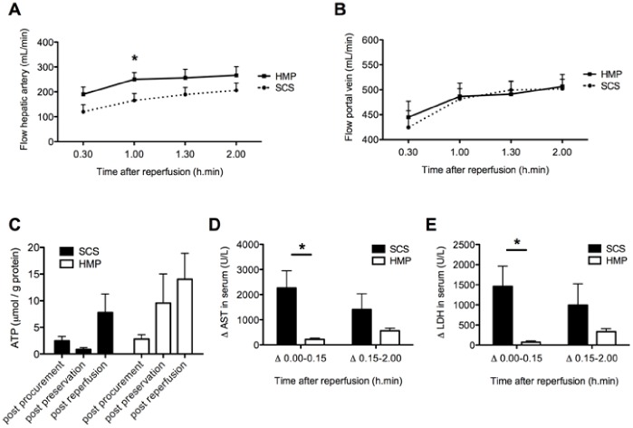
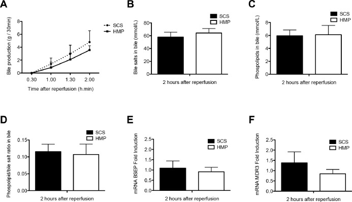
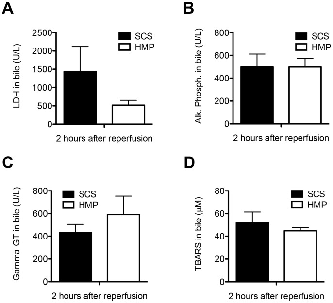
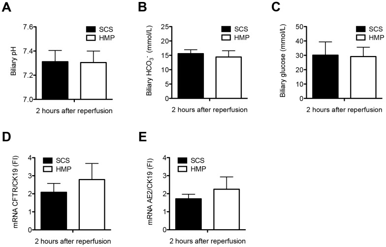
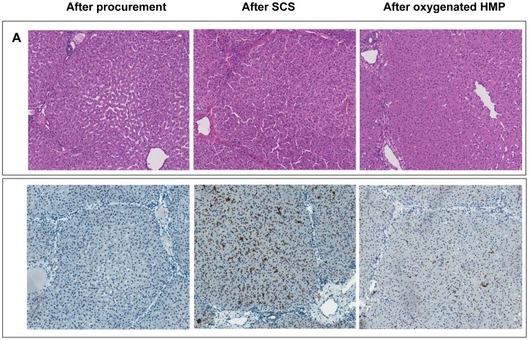
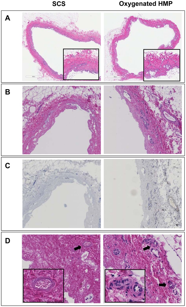
Results: After reperfusion, arterial flow was higher in the HMP group, compared to SCS (251±28 vs 166±28 mL/min, respectively, after 1 hour of reperfusion; p = 0.003). Release of hepatocellular enzymes was significantly higher in the SCS group. Markers of biliary epithelial injury (biliary LDH, gamma-GT) and function (biliary pH and bicarbonate, and biliary transporter expression) were similar in the two groups. However, histology of bile ducts revealed significantly less arteriolonecrosis of the peribiliary vascular plexus in HMP preserved livers (>50% arteriolonecrosis was observed in 7 bile ducts of the SCS preserved livers versus only 1 bile duct of the HMP preserved livers; p = 0.024).

Conclusions: Oxygenated HMP prevents arteriolonecrosis of the peribiliary vascular plexus of the bile ducts of DCD pig livers and results in higher arterial flow after reperfusion. Together this may contribute to better perfusion of the bile ducts, providing a potential advantage in the post-ischemic recovery of bile ducts.


Conflict of interest statement

Competing Interests: The authors have declared that no competing interests exist.

Figures

Figure 1
Figure 1. Perfusion characteristics during and changes in serum concentrations of AST and LDH after 2 h of normothermic ex vivo sanguineous reperfusion of DCD livers that were preserved by either 4 h of oxygenated HMP or SCS.
Panel A: Blood flow through the hepatic artery. Panel B: Blood flow through the portal vein. Panel C: Changes in hepatic energy content as reflected by hepatic ATP content. Panel D and E: Relative increase of liver enzymes was significantly greater for SCS preserved livers, compared to livers preserved by oxygenated HMP. *p-value <0.05.
Figure 2
Figure 2. Parameters of hepatocellular secretory function of DCD livers that were preserved by either 4 h of oxygenated HMP or SCS and subsequently reperfused for 2 h by normothermic ex vivo sanguineous perfusion.
Panel A: Evolution of bile production in the HMP and SCS group. Panel B and C: Biliary concentration of bile salts and phospholipids, respectively. Panel D: Bile salt toxicity, as represented by the ratio of biliary bile salt and phospholipid concentrations. There were no statistically significant differences between the groups. Panel E–F: Relative mRNA expression of the main hepatocellular bile transporters BSEP (bile salt export pump; Abcb11) and MDR3 (multidrug resistance protein 3, Abcb4) after 2 h of ex vivo sanguineous reperfusion of DCD livers that were preserved by either 4 h of oxygenated HMP or SCS. There were no significant differences between the two groups.
Figure 3
Figure 3. Comparison of biochemical parameters of bile duct injury and oxidative stress in DCD livers that were preserved by either 4 h of oxygenated HMP or 4 h of SCS and subsequently reperfused for 2 h by normothermic ex vivo sanguineous perfusion.
Panel A–C: Concentration of LDH, alkaline phosphatase, and gamma-GT in bile samples at 2 h after reperfusion. Panel D: Comparison of biliary concentration of TBARS, a marker for oxidative stress and lipid peroxidation in bile ducts, at 2 h after graft reperfusion. There were no significant differences between the SCS preserved and HMP preserved livers.
Figure 4
Figure 4. Comparison of functional parameters of biliary epithelial cell function in DCD livers that were preserved by either 4 h of oxygenated HMP or 4 h of SCS and subsequently reperfused for 2 h by normothermic ex vivo sanguineous perfusion.
There were no significant differences in biliary pH (Panel A), biliary bicarbonate (Panel B) and glucose concentration (Panel C) at 2 h after reperfusion between the two groups. Relative mRNA expression of the main cholangiocyte transporter proteins involved in biliary bicarbonate secretion, CFTR (cystic fibrosis transmembrane conductance regulator; ABC35) (Panel D) and AE2 (anion exchanger 2; SLC4A2) (Panel E) after 2 h of ex vivo sanguineous reperfusion of DCD livers that were preserved by either 4 h of oxygenated HMP or SCS. There were no significant differences between the two groups.
Figure 5
Figure 5. Representative examples of histology of liver parenchyma of DCD liver grafts preserved by either 4 h of SCS or 4 h of oxygenated HMP followed by 2 h by normothermic ex vivo sanguineous perfusion.
Panel A: H&E staining of a central biopsy of the liver parenchyma. Panel B: Caspase-3 immunohistochemistry of liver parenchyma showing less intense caspase-3 staining of hepatocytes, sinusoidal endothelial cells, and Kupffer cells in the HMP group, compared to the SCS group. Brown color indicates immunopositivity. Original magnification 200x.
Figure 6
Figure 6. Representative examples of H&E histology of bile ducts of DCD liver grafts preserved by either 4 h of oxygenated HMP or 4 h of SCS followed by 2 h by normothermic ex vivo sanguineous perfusion.
Panel A: extrahepatic bile duct immediately after procurement (magnification 200x). The insert represents a higher magnification of 400x. Panel B: extrahepatic bile duct after preservation and reperfusion (magnification 280x). Panel C: Immunohistochemistry for activated caspase-3 of the same bile ducts as presented in panel B (brown color indicates immunopositivity; counterstaining with hematoxylin). Very few caspase-3 positive cells were detected in the bile duct wall stroma. Remnant biliary epithelial cells (i.e. in the peribiliary glands) were not positive for acitivated caspase-3. Panel D: higher magnification (400x) of extrahepatic bile ducts focusing on the peribiliary plexus. Arrows indicate peribiliary arterioles. The insert represents a higher magnification of the peribiliary arterioles (800x). Bile ducts of livers preserved by oxygenated HMP displayed significantly less signs of arteriolonecrosis, compared to livers preserved by SCS (see also Table 1 ).

References

    1. Pine JK, Aldouri A, Young AL, Davies MH, Attia M, et al. (2009) Liver transplantation following donation after cardiac death: An analysis using matched pairs. Liver Transpl 15: 1072–1082. - PubMed
    1. Meurisse N, Vanden Bussche S, Jochmans I, Francois J, Desschans B, et al. (2012) Outcomes of liver transplantations using donations after circulatory death: A single-center experience. Transplant Proc 44: 2868–2873. - PubMed
    1. Suarez F, Otero A, Solla M, Arnal F, Lorenzo MJ, et al. (2008) Biliary complications after liver transplantation from maastricht category-2 non-heart-beating donors. Transplantation 85: 9–14. - PubMed
    1. Dubbeld J, Hoekstra H, Farid W, Ringers J, Porte RJ, et al. (2010) Similar liver transplantation survival with selected cardiac death donors and brain death donors. Br J Surg 97: 744–753. - PubMed
    1. Verdonk RC, Buis CI, van der Jagt EJ, Gouw AS, Limburg AJ, et al. (2007) Nonanastomotic biliary strictures after liver transplantation, part 2: Management, outcome, and risk factors for disease progression. Liver Transpl 13: 725–732. - PubMed

Publication types

MeSH terms