. 2014 Feb 13;9(2):e88772.
doi: 10.1371/journal.pone.0088772. eCollection 2014.

Fibroblast activation protein (FAP) is essential for the migration of bone marrow mesenchymal stem cells through RhoA activation

Affiliations

Fibroblast activation protein (FAP) is essential for the migration of bone marrow mesenchymal stem cells through RhoA activation

Kuei-Min Chung et al. PLoS One. .

Abstract

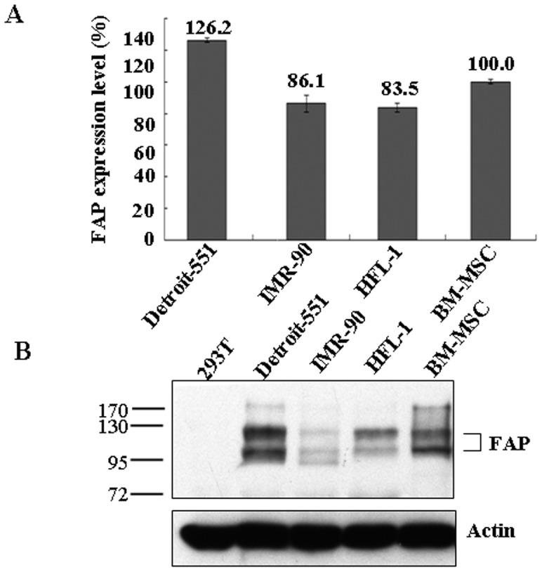
Background: The ability of human bone marrow mesenchymal stem cells (BM-MSCs) to migrate and localize specifically to injured tissues is central in developing therapeutic strategies for tissue repair and regeneration. Fibroblast activation protein (FAP) is a cell surface serine protease expressed at sites of tissue remodeling during embryonic development. It is also expressed in BM-MSCs, but not in normal tissues or cells. The function of FAP in BM-MSCs is not known.

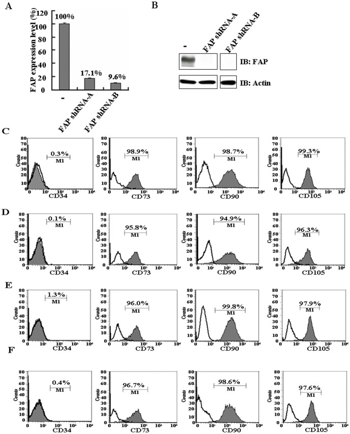
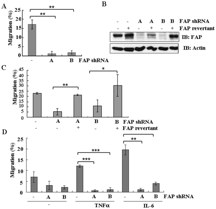
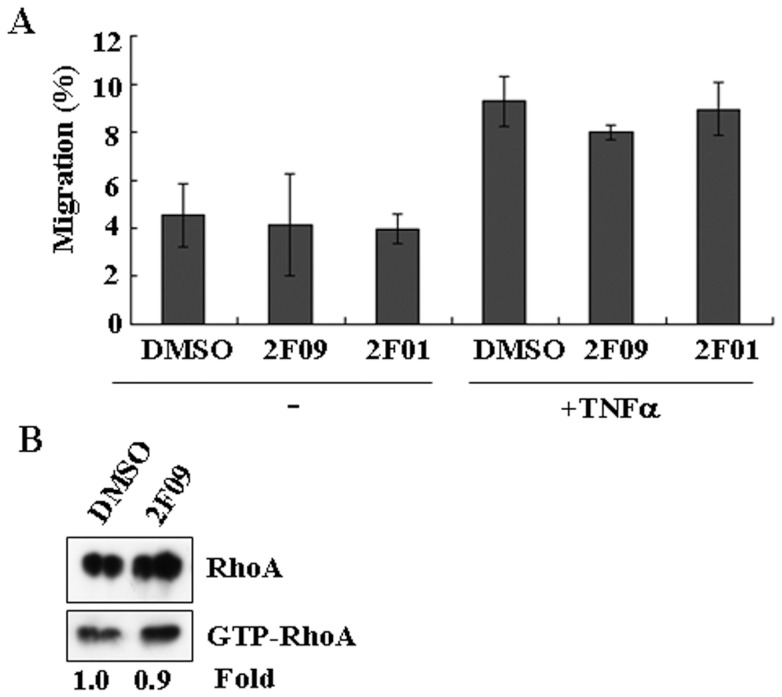
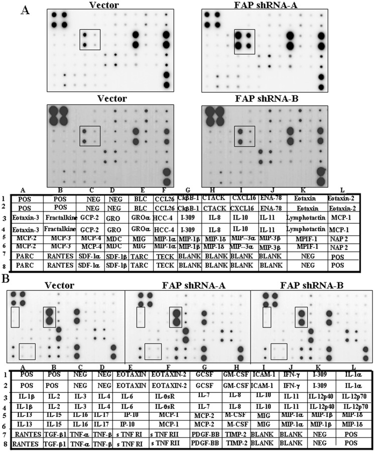
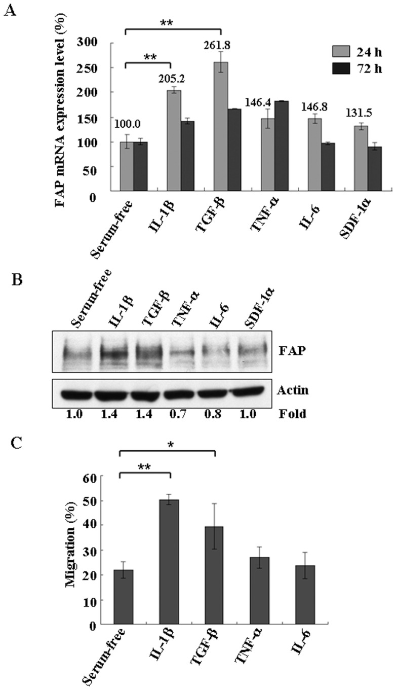
Principal findings: We found that depletion of FAP proteins significantly inhibited the migration of BM-MSCs in a transwell chemotaxis assay. Such impaired migration ability of BM-MSCs could be rescued by re-expressing FAP in these cells. We then demonstrated that depletion of FAP activated intracellular RhoA GTPase. Consistently, inhibition of RhoA activity using a RhoA inhibitor rescued its migration ability. Inhibition of FAP activity with an FAP-specific inhibitor did not affect the activation of RhoA or the migration of BM-MSCs. Furthermore, the inflammatory cytokines interleukin-1beta (IL-1β) and transforming growth factor-beta (TGF-β) upregulated FAP expression, which coincided with better BM-MSC migration.

Conclusions: Our results indicate FAP plays an important role in the migration of BM-MSCs through modulation of RhoA GTPase activity. The peptidase activity of FAP is not essential for such migration. Cytokines IL-1β and TGF-β upregulate the expression level of FAP and thus enhance BM-MSC migration.


Conflict of interest statement

Competing Interests: The authors have declared that no competing interests exist.

Figures

Figure 1
Figure 1. Characterization of FAP expression level in BM-MSCs.
(A) Analysis of the mRNA expression levels of FAP in different human cell lines as indicated. Quantification was carried out by qRT–PCR as described in the Materials and methods section. The error bars are showing standard deviation (SD). (B) Expression of FAP detected by western blot analysis with anti-FAP antibodies. Immunoblotting with anti-actin antibodies was used as a loading control.
Figure 2
Figure 2. FAP depletion in BM-MSCs by lentiviral infection.
(A) BM-MSCs were depleted of FAP by transfection with lentiviruses encoding two FAP shRNA sequences. The efficiency of shRNA-based downregulation was determined by qRT–PCR. Results are given as the percentage of change in mRNA expression relative to pLKO empty vector-infected cells set as 100%. The error bars are showing standard deviation (SD). (B) The efficiency of shRNA-based downregulation was determined by western blot analysis. Data are representative of three independent experiments. (C–F) Flow cytometric analysis of cell surface markers on FAP-depleted BM-MSCs. (C) Parental BM-MSCs were analyzed using antibodies against CD34, CD73, CD90, and CD105. (D) Vector infected BM-MSCs were analyzed using antibodies against CD34, CD73, CD90, and CD105. (E) As in (D) but with FAP shRNA-A infected BM-MSCs. (F) As in (D) but with FAP shRNA-B infected BM-MSCs.
Figure 3
Figure 3. FAP depletion and the migration of BM-MSCs.
(A) BM-MSCs were infected by lentiviruses encoding FAP shRNA-A (A), shRNA-B (B), or vector control (–). The migration ability of FAP-depleted BM-MSCs in the presence of 10% FBS was analyzed by Boyden chamber transwell assays in vitro. Cells were seeded in the upper wells of the chambers in IMDM and the lower wells contained the same medium with 10% FBS. Cells were allowed to migrate for 48 h and the numbers of cells that migrated were determined as described in the Materials and methods section. Data are representative of three separate experiments each performed in triplicate. The error bars are showing standard deviation (SD). (B) FAP-depleted BM-MSCs were infected with lentiviruses expressing an FAP revertant. The protein expression level of FAP revertant in FAP knockdown BM-MSCs was determined by western blot analysis. (C) Results are shown as in (A) but with the FAP-depleted cells infected with a lentivirus expressing an FAP revertant. Data are representative of three separate experiments each performed in triplicate. The error bars are showing standard deviation (SD). (D) Results are shown as in (A) but the lower wells of the transwell chambers contained IMDM with TNF-α (100 ng/ml) or IL-6 (100 ng/ml). Data are representative of three separate experiments each performed in triplicate. Means and standard deviations are given in each case. The error bars are showing standard deviation (SD). The statistically significant differences between the groups were assessed using a two-tailed Student's t test. The degree of significance is indicated as fellows: *p<0.05; **p<0.001; ***p<0.0001.
Figure 4
Figure 4. Rho GTPase activation in FAP-depleted BM-MSCs.
(A) The amount of GTP-bound RhoA was determined with the cells depleted of FAP, as described in the Materials and methods section. The amounts of GTP-bound RhoA were normalized against total RhoA protein present in cell lysates and expressed as fold induction compared with the cells infected with the vector control. Data are representative of two independent experiments. (B) Results are shown as in (A) except that GTP-bound Rac1 was used. Data are representative of two independent experiments. (C) Effect of RhoA inhibitors on the migration of FAP-depleted BM-MSCs was determined by transwell assays. FAP-depleted BM-MSCs were pretreated without or with the RhoA inhibitor C2I-C3 (1 µg/ml) (Cytoskeleton Inc.) in IMDM for 2 h and seeded in the upper wells of the chambers. The lower wells contained IMDM with 10% FBS. Cells were allowed to migrate for 48 h and absolute cell numbers were determined as described in the Materials and methods section. Data are representative of three separate experiments each performed in triplicate. Means and standard deviations were calculated. The error bars are showing standard deviation (SD). The statistically significant differences between the groups were assessed using a two-tailed Student's t test. The degree of significance is indicated as fellows: **p<0.001; ***p<0.0001.
Figure 5
Figure 5. Inhibition of FAP activity had no effect on BM-MSC migration.
(A) BM-MSCs were incubated with DMSO, 2F09 (10 µM) or 2F01 (10 µM) in IMDM before being seeded in the upper wells of transwell chambers. The lower wells contained IMDM with TNF-α (100 ng/ml). Cells were allowed to migrate for 48 h and absolute cell numbers were determined as described in the Materials and methods section. Data are representative of three separate experiments each performed in triplicate. The error bars are showing standard deviation (SD). (B) Results are shown as in (A) except that the cells were incubated with dimethyl sulfoxide (DMSO) solvent or 2F09 (10 µM) in IMDM for 2 h before lysis to determine the amount of GTP-bound RhoA as described in the Materials and methods section. Data are representative of two independent experiments.
Figure 6
Figure 6. Effect of FAP depletion on chemokine and inflammatory cytokine secretion profiles.
(A) The protein levels of chemokines in FAP-depleted BM-MSCs were compared to the control with RayBio human chemokine antibody array 1, according to the manufacturer's manual. The corresponding chemokines spotted on the membrane was shown below the blot. The highlighted box in the blot indicated the position of GRO and GRO-α. Data are representative of two independent experiments. (B) As in (A) but with RayBio human inflammation antibody array 3. The highlighted box in the blot indicated the position of cytokines, IL-1β, IL-6, TGF-β and TNF-α, which are known to involve in cellular movement. Representative results of two independent experiments.
Figure 7
Figure 7. IL-1β and TGF-β increased the expression of FAP in BM-MSCs.
(A) BM-MSCs were incubated with IL-1β (100 ng/ml), TGF-β (100 ng/ml), TNF-α (100 ng/ml), IL-6 (100 ng/ml), or SDF-1α (100 ng/ml) under serum-free conditions in IMDM for 24 or 72 h. The cells were harvested and lysed to determine the mRNA levels of FAP by qRT–PCR. Results are given as the percentage change in mRNA expression relative to untreated cells, which was set at 100%. Data are representative of three independent experiments. The error bars are showing standard deviation (SD). (B) Results are as shown in (A) except that the protein expression level of FAP was determined after incubation for 48 h with the indicated cytokines/chemokines in IMDM. Immunoblotting was carried out with anti-FAP antibodies. Immunoblotting for actin was used as a loading control. Data are representative of three independent experiments. (C) The effects of IL-1β and TGF-β on the migration of BM-MSCs were determined by transwell assays. BM-MSCs were incubated without or with IL-1β (100 ng/ml), TGF-β (100 ng/ml), TNF-α (100 ng/ml) and IL-6 (100 ng/ml) grown under serum-free conditions in IMDM for 48 h before being seeded in the upper wells of transwell chambers. The lower wells contained TNF-α (100 ng/ml) in IMDM to stimulate migration. Cells were allowed to migrate for 24 h and absolute cell numbers were determined as described in the Materials and methods section. Data are representative of three separate experiments each performed in triplicate. Means and standard deviations were calculated. The error bars are showing standard deviation (SD). The statistically significant differences between the groups were assessed using a two-tailed Student's t test. The degree of significance is indicated as fellows: *p<0.05; **p<0.001.

Similar articles

Cited by

References

    1. Rosenblum JS, Kozarich JW (2003) Prolyl peptidases: a serine protease subfamily with high potential for drug discovery. Curr Opin Chem Biol 7: 496–504. - PubMed
    1. Scanlan MJ, Raj BK, Calvo B, Garin-Chesa P, Sanz-Moncasi MP, et al. (1994) Molecular cloning of fibroblast activation protein alpha, a member of the serine protease family selectively expressed in stromal fibroblasts of epithelial cancers. Proc Natl Acad Sci U S A 91: 5657–5661. - PMC - PubMed
    1. Rettig WJ, Garin-Chesa P, Beresford HR, Oettgen HF, Melamed MR, et al. (1988) Cell-surface glycoproteins of human sarcomas: differential expression in normal and malignant tissues and cultured cells. Proc Natl Acad Sci U S A 85: 3110–3114. - PMC - PubMed
    1. Huber MA, Kraut N, Park JE, Schubert RD, Rettig WJ, et al. (2003) Fibroblast activation protein: differential expression and serine protease activity in reactive stromal fibroblasts of melanocytic skin tumors. J Invest Dermatol 120: 182–188. - PubMed
    1. Chen WT, Kelly T (2003) Seprase complexes in cellular invasiveness. Cancer Metastasis Rev 22: 259–269. - PubMed

Publication types

MeSH terms

LinkOut - more resources