. 2014 Feb 14;9(2):e88797.
doi: 10.1371/journal.pone.0088797. eCollection 2014.

Systemic delivery of siRNA down regulates brain prion protein and ameliorates neuropathology in prion disorder

Affiliations

Systemic delivery of siRNA down regulates brain prion protein and ameliorates neuropathology in prion disorder

Sylvain Lehmann et al. PLoS One. .

Abstract

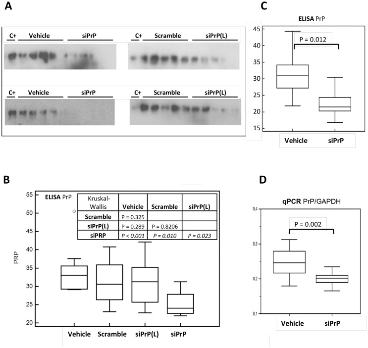
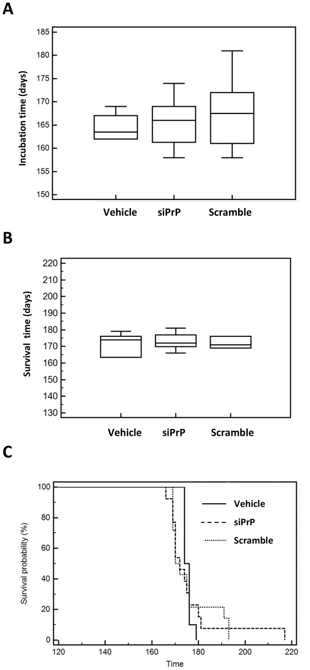
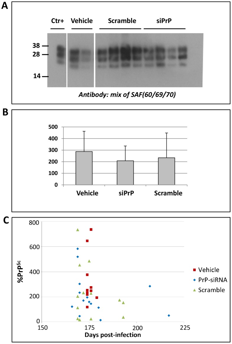
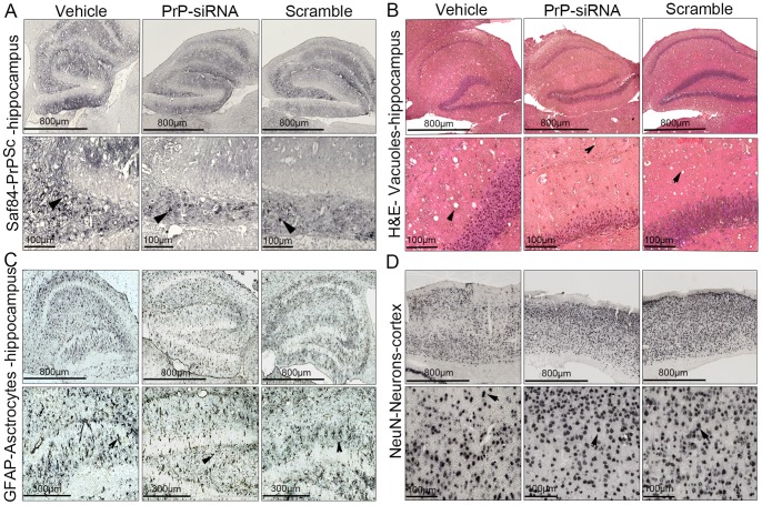
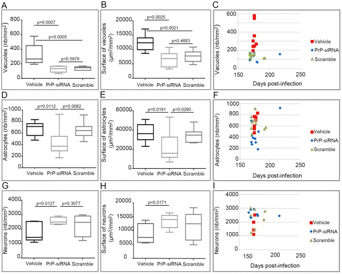
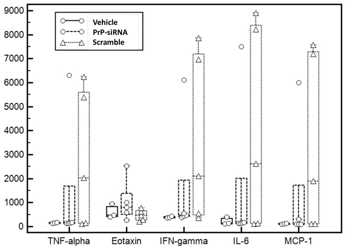
One of the main challenges for neurodegenerative disorders that are principally incurable is the development of new therapeutic strategies, which raises important medical, scientific and societal issues. Creutzfeldt-Jakob diseases are rare neurodegenerative fatal disorders which today remain incurable. The objective of this study was to evaluate the efficacy of the down-regulation of the prion protein (PrP) expression using siRNA delivered by, a water-in-oil microemulsion, as a therapeutic candidate in a preclinical study. After 12 days rectal mucosa administration of Aonys/PrP-siRNA in mice, we observed a decrease of about 28% of the brain PrP(C) level. The effect of Aonys/PrP-siRNA was then evaluated on prion infected mice. Several mice presented a delay in the incubation and survival time compared to the control groups and a significant impact was observed on astrocyte reaction and neuronal survival in the PrP-siRNA treated groups. These results suggest that a new therapeutic scheme based an innovative delivery system of PrP-siRNA can be envisioned in prion disorders.


Conflict of interest statement

Competing Interests: The author have read the journal’s policy and have the following conflicts: Medesis Pharma provided financial support for this collaborative research project. Sarah Resina, Elsa Brillaud, Charles Vincent, Sophie Poupeau, Jean-Claude Maurel and Patrick Maurel are employees of Medesis Pharma. Medesis Pharma was involved in experimental work and collection of raw data. Data analysis and writing of the article and conclusion were performed by the academic authors who have no link with Medesis Pharma. This does not alter our adherence to all the PLOS ONE policies on sharing data and materials.

Figures

Figure 1
Figure 1. In vivo PrPC expression in AONYS/siRNA-treated mice.
Panel A. Brain homogenates of mice treated for 12(see Table 1) with two doses of PrPC siRNA (Aonys/siPrP 300 (L) and 600 µg/kg), the vehicle only and scrambled-siRNA, were analysed for PrPC accumulation by Western blot. The C+ lane corresponds to a brain homogenate of untreated mice. A decrease of PrPC was apparent in siPrP treated mice. However there was a relative variability in detection in relation with the low linearity of the western blot. Panel B. Samples in A were analysed using ELISA for quantitation of PrPC. Graphic results of normalized PrPC were presented as medians and interquartile ranges. Statistical analysis (Kruskal-Wallis test) was used to evaluate the significance of the difference between the groups. The siPrP group (600 µg/kg) presents a significant reduction of PrPC levels. Panels C and D. Additional experiments were carried out (10 mice/groups) in which mice were treated or not with Aonys/PrPC siRNA at 600 µg/kg. Panel C. PrPC ELISA. Panel D. RT-Q-PCR quantitation (light Cycler) of PrP and GAPDH in Graphic results were presented as medians and interquartile ranges. Statistical analysis (Kruskal-Wallis test) was used to evaluate the significance of the difference between the groups. The siPrP group presents a significant reduction of both PrPC protein and RNA levels.
Figure 2
Figure 2. Impact of siRNA on incubation and survival time.
Panel A. The incubation time in scrapie infected mice based on the presence of at least three clinical signs (among: waddling gait, flattened back, rough coat, sticky eye discharge, weight loss, very jumpy, hunched, incontinence) was plotted as medians and interquartile ranges in the three groups: Aonys/PrPC siRNA (siPrP 600 µg/kg), Aonys vehicle only and Aonys/scrambled-siRNA. The survival times of the three groups of infected mice were plotted as medians and interquartile ranges (Panel B.) and Kaplan-Meier survival curves (Panel C.). No statistical difference was observed between each group neither for the incubation period nor the survival time.
Figure 3
Figure 3. Impact of siRNA on brain PrPSc content.
Western blot analysis of PrPSc in mouse brain lysates (Panel A.). Only representative samples of the mouse population are presented here. The classical ME7 PrPSc bands were present at various intensity levels. The Ctr+ lane corresponds to a brain homogenate of ME7 scrapie mice loaded as a standard in every blot. Quantitation of the signal (Panel B.) normalized in each blot with respect to the Ctr+ sample showed no significant difference between groups. (Panel C.) Individual PrPSc levels and survival times.
Figure 4
Figure 4. Histological and Immunohistolochemical illustration.
Panel A. PrPSc immunostaining was observed in all the mice (arrow). Panel B. Vacuolar lesions revealed after Hematoxylin & Eosin staining were apparent in all conditions. Immunohistochemical detections of the GFAP astrocyte marker (Panel C) and the neuronal marker NeuN were also performed (Panel D).
Figure 5
Figure 5. Histological and Immunohistolochemical data analysis.
Panels A,B. Quantitation of vacuolar lesions after Hematoxylin & Eosin staining (figure 4) revealed that siRNA have a slight impact. Panels D,E. Quantitation of astrocyte based on the GFAP immunostaining (figure 4) showed significant differences between the Aonys/PrP-siRNA treated group and the two control groups (vehicle and scrambled-siRNA). Panels G,H. Quantitation of neurons based on the neuronal marker NeuN (figure 4) revealed statistical differences between mice treated with PrP-siRNA and vehicle. Panels C,F,I. Data of individual levels of vacuolisation, NeuN or GFAP counts as a function of survival time did not show any correlation.
Figure 6
Figure 6. Cytokines analysis.
Graphic representation plotted as medians and interquartile ranges of selected blood cytokine levels in the three mice groups (see table 2 for full data). Note that outliners characterized by very high cytokine levels were present in the siRNA groups only.

References

    1. Sikorska B, Liberski PP (2012) Human prion diseases: from kuru to variant creutzfeldt-jakob disease. Subcell Biochem 65: 457–496. - PubMed
    1. Parchi P, Saverioni D (2012) Molecular pathology, classification, and diagnosis of sporadic human prion disease variants. Folia Neuropathol 50: 20–45. - PubMed
    1. Prusiner SB (1982) Novel proteinaceous infectious particles cause scrapie. Science 216: 136–144. - PubMed
    1. Bueler H, Aguzzi A, Sailer A, Greiner RA, Autenried P, et al. (1993) Mice devoid of PrP are resistant to scrapie. Cell 73: 1339–1347. - PubMed
    1. Sailer A, Bueler H, Fischer M, Aguzzi A, Weissmann C (1994) No propagation of prions in mice devoid of PrP. Cell 77: 967–968. - PubMed

Publication types