. 2014 Feb 13;9(2):e89009.
doi: 10.1371/journal.pone.0089009. eCollection 2014.

High-dose irradiation induces cell cycle arrest, apoptosis, and developmental defects during Drosophila oogenesis

Affiliations

High-dose irradiation induces cell cycle arrest, apoptosis, and developmental defects during Drosophila oogenesis

Hee Jin Shim et al. PLoS One. .

Abstract

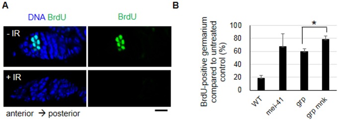
Ionizing radiation (IR) treatment induces a DNA damage response, including cell cycle arrest, DNA repair, and apoptosis in metazoan somatic cells. Because little has been reported in germline cells, we performed a temporal analysis of the DNA damage response utilizing Drosophila oogenesis as a model system. Oogenesis in the adult Drosophila female begins with the generation of 16-cell cyst by four mitotic divisions of a cystoblast derived from the germline stem cells. We found that high-dose irradiation induced S and G2 arrests in these mitotically dividing germline cells in a grp/Chk1- and mnk/Chk2-dependent manner. However, the upstream kinase mei-41, Drosophila ATR ortholog, was required for the S-phase checkpoint but not for the G2 arrest. As in somatic cells, mnk/Chk2 and dp53 were required for the major cell death observed in early oogenesis when oocyte selection and meiotic recombination occurs. Similar to the unscheduled DNA double-strand breaks (DSBs) generated from defective repair during meiotic recombination, IR-induced DSBs produced developmental defects affecting the spherical morphology of meiotic chromosomes and dorsal-ventral patterning. Moreover, various morphological abnormalities in the ovary were detected after irradiation. Most of the IR-induced defects observed in oogenesis were reversible and were restored between 24 and 96 h after irradiation. These defects in oogenesis severely reduced daily egg production and the hatch rate of the embryos of irradiated female. In summary, irradiated germline cells induced DSBs, cell cycle arrest, apoptosis, and developmental defects resulting in reduction of egg production and defective embryogenesis.


Conflict of interest statement

Competing Interests: The authors have declared that no competing interests exist.

Figures

Figure 1
Figure 1. IR-induced G2 arrest in mitotically dividing germline and somatic cells in wild type and checkpoint mutant ovaries.
The wild type or indicated mutant adult females were irradiated with 0 or 40 Gy and the ovaries were dissected at the indicated time points. The ovaries were stained with an anti-Orb (green) and anti-phospho-histone H3 (PH3) antibody (red) to detect germline cells in region 2/3 and mitotic cells, respectively. The DNA was stained with DAPI (blue). (A) Representative images of the wild type germarium containing PH3-stained germline cells (arrow) or somatic follicle cells (arrowhead) are shown in the absence of irradiation (-IR) or 1 h after irradiation (+IR). Germariums are oriented as the anterior to the top. Bar, 5 µm. (B) The wild type ovaries were dissected 1, 6, 8, 24, and 72 h after 40 Gy of irradiation and stained with PH3. The number of germariums containing PH3-positive germline cells in region 1 and somatic cells in region 3 were counted. The values represent the percentage of germarium containing PH3-positive germline cells (solid line) and somatic cells (dotted line). The data are presented as the mean and standard deviation of three independent experiments. The total number of germarium for each time point was at least 150. (C) The ovaries of checkpoint mutant adult females were stained with PH3 1 h after 40 Gy. The germariums containing PH3-stained germline cells (black bar) or somatic cells (gray bar) were counted. The values represent the relative percentage of PH3-positive germariums in irradiated ovaries compared to the untreated controls. The data are presented as the mean and standard deviation of three independent experiments. The total number of germariums for each genotype was at least 150. * p<0.05 (the grp mutant versus the grp mnk double mutant).
Figure 2
Figure 2. IR-induced S-phase checkpoint in mitotically dividing germline cells in wild type and checkpoint mutant females.
The BrdU incorporation assay was performed in wild type and checkpoint mutant ovaries 1(A) Representative images of a wild type germarium assayed for BrdU incorporation (green) in the germline cells before and after irradiation are shown. The DNA was stained with DAPI (blue). Bar, 10 µm. (B) The number of germariums containing a strong BrdU signal in region 1 were counted. The values represent the relative percentage of germarium containing BrdU-positive germline cells in irradiated samples compared to the untreated controls. The data are presented as the means and standard deviations of three independent experiments. The total number of germarium for each cell type at different time points were at least 220. * p = 0.0016 (the grp mutant versus the grp mnk double mutant).
Figure 3
Figure 3. IR-induced apoptosis during oogenesis.
The wild type or mutant adult females were irradiated at 40 Gy, and the ovaries were stained by using the TUNEL method to detect the apoptotic cells. (A) The values represent the percentage of wild type or the indicated mutant ovarioles containing TUNEL-positive germline cells in region 2 at 6 h after irradiation. The data are presented as the mean and standard deviation of two independent experiments. The total number of ovarioles counted for each genotype was at least 100. * p = 0.085. ** p = 0.21. (B) The values represent the percentage of wild type ovarioles containing TUNEL-positive germline cells in region 2 (solid line) or degenerating egg chambers at stages 7-10 (dotted line) at 0, 6, 24, 48, 72, and 96 h after irradiation. The data are presented as the mean and standard deviation of two independent experiments. The total number of ovarioles counted at each time point was at least 70. (C) The values represent the percentage of wild type egg chambers at stages 2–6 or 7–10 containing more than 5 TUNEL-positive follicle cells 6 h after irradiation. The total number of egg chambers counted at each time point was at least 150.
Figure 4
Figure 4. Generation and repair of DSBs detected by H2Av phosphorylation in oocytes after IR treatment.
The wild type females were irradiated at 40 Gy, and the ovaries were stained for γH2Av (red), DNA (blue), and Orb (green). (A) Representative confocal images of ovariole before and 1 h after irradiation are shown. Bar, 50 µm. (B) Representative confocal images of stage 3 egg chambers 0, 1, and 8 h after irradiation are shown. The oocytes containing a high level of Orb proteins are indicated by a white circle. Bar, 5 µm. (C) The percentage of oocytes at stage 3 stained with γH2Av was determined at 0, 1, 8, 24, 48, and 72 h after irradiation. The data are presented as the mean and standard deviation of two independent experiments. The total number of oocytes at each time point was at least 14.
Figure 5
Figure 5. Meiotic checkpoint and defective eggshell induced by IR treatment.
(A–B) Karyosome defects after IR treatment. Wild type females were irradiated at 40 Gy, and the ovaries were stained for DNA with propidium iodide (red) and for oocyte cytoplasm with an anti-Orb antibody (green). (A) Representative confocal images of stage 6 egg chambers before (a) and after irradiation (b, c, and d) are shown. Bar, 10 µm. The nuclei of oocytes are indicated with a white arrow. (B) The percentage of oocytes at stage 6 with defective karyosome was counted at 0, 1, 8, 24, 48, and 72 h after irradiation. The morphology of the karyosome similar to (Ad) was considered as defective. The data are presented as the mean and standard deviation of at least two independent experiments. The total number of oocytes at each time point was at least 21. (C–D) Dorsal-ventral patterning defects of the embryos laid by the irradiated females. (C) Representative images of a normal embryo from an untreated female (-IR) and a defective embryo from a wild type female irradiated at 40 Gy (+IR) are shown. (D) Wild type or mnk mutant females were irradiated at 40 Gy and the embryos were collected every 24 h. The percentage of embryos with dorsal-ventral patterning defects were counted each day. Day 1 corresponds to the embryos collected 0–24 h after irradiation. The data are presented as the mean and standard deviation of at least two independent experiments. At least 100 embryos were observed for each time point in each experiment. (E–F) Eggshell defects in the embryos derived from irradiated females. (E) The embryos derived from the untreated (-IR, normal eggshell) and 40 Gy irradiated (+IR) females are shown. The embryos from irradiated female exhibited irregular eggshells (second panel, indicated in the red dotted line) or lucid eggshells (third panel). (F) The embryos were collected from the irradiated females every 24 h, and the percentages of the embryos with thin eggshell phenotypes were counted. Day 1 represents the embryos collected at the first 24 h after irradiation. The data are presented as the mean and standard deviation of three independent experiments. At least 100 embryos were observed for each time point.
Figure 6
Figure 6. Morphological defects of ovarioles induced by high-dose irradiation.
The wild type females were irradiated at 40 Gy, and the germline cells were stained with an anti-Vasa antibody (green) and DAPI (blue) each day after irradiation. (A) Various defects (indicated by an arrow) were observed in oogenesis after irradiation. Representative images exhibiting the following defects are shown: germarium lacking anti-Vasa-stained germline cells in region 3 and/or region 2b (a), in region 1 (b), or in the entire germarium (c); ovarioles lacking a couple of early stage egg chambers (j) and several late stage egg chambers (k); germarium exhibiting fusion of two cysts (d); fused egg chambers (g); single germline cells in the germarium (e); abnormal number of germline cells in the egg chambers (f); ovarioles containing smaller egg chambers at more posterior positions (g, arrowhead); egg chambers with an abnormal shape (h); and egg chambers containing mislocalized nurse cells (l). The germarium (g) and the stage of the egg chambers are shown in (j) and (k). The scale bars represent 20 µm (a–i) and 50 µm (j–l). (B) Irradiation induced loss of germline cells in the germarium. The percentage of germarium lacking anti-Vasa-stained germline cells in region 3 and/or 2 (blue dots), region 1 (red diamonds), or the entire germarium (green triangles) was counted each day after irradiation. The values represent the means and standard deviations from two independent experiments. At least 55 ovarioles were counted in total for each time point. (C) Morphological defects observed in the germarium and egg chambers. The percentage of germarium containing morphological defects in the germline cells (blue dots) or extracellular materials (green triangles) was counted. The percentage of ovarioles containing an abnormal egg chamber morphology (red diamonds) was also calculated. The values represent the means and standard deviations from two independent experiments. At least 55 ovarioles were counted in total for each time point.
Figure 7
Figure 7. Fecundity, fertility, and the development of embryos derived from irradiated adult females.
(A–C) Fecundity and fertility of the wild type irradiated females. Two- to three-day-old adult females were irradiated with 0 or 40 Gy and the embryos were collected every 24 h. Day 1 corresponds to the egg collection between 0 and 24 h. The number of embryos per female (A) and the hatch rate of these embryos (B) were determined. Two independent experiments were performed using at least 5 cages for each treatment in the experiment, and the cage contained 3 pairs of females and males. The data are presented as the mean and standard deviation of the number of embryos per female and the hatch rate of those embryos was calculated from each cage. The grey X in (A) represents the percentage of egg production by irradiated females compared to untreated females. (C) The number of hatched larvae derived from one female was calculated and shown as above. (D–F) Development of the first instar larvae produced from irradiated females. (D–E) First instar larvae (n = 100) were obtained from the daily collection of eggs laid by the females treated with 0 or 40 Gy, and the number of pupae and eclosed adults was counted. The values represent the percentage of the total number of pupae formed (D) and the percentage of total number of adults generated from the pupae (E). The data are presented as the mean and standard deviation of three independent experiments. (F) The Minute phenotypes were observed in the adult females produced from females treated with 0 or 40 Gy. The percentage of adults with defective bristles is shown. The data are presented as the mean and standard deviation of a total of 42 females observed in three independent experiments.

Similar articles

Cited by

References

    1. Song YH (2005) Drosophila melanogaster: a model for the study of DNA damage checkpoint response. Mol Cells 19: 167–179. - PubMed
    1. Touchberry RW, Defries JC (1964) Genetic Effects of Gamma-Irradiation on Egg Production and Adult Emergence of Drosophila Melanogaster. Genetics 49: 387–400. - PMC - PubMed
    1. Drummond-Barbosa D, Spradling AC (2001) Stem cells and their progeny respond to nutritional changes during Drosophila oogenesis. Dev Biol 231: 265–278. - PubMed
    1. Abdu U, Brodsky M, Schupbach T (2002) Activation of a meiotic checkpoint during Drosophila oogenesis regulates the translation of Gurken through Chk2/Mnk. Curr Biol 12: 1645–1651. - PubMed
    1. Sibon OC, Laurencon A, Hawley R, Theurkauf WE (1999) The Drosophila ATM homologue Mei-41 has an essential checkpoint function at the midblastula transition. Curr Biol 9: 302–312. - PubMed

Publication types

MeSH terms

LinkOut - more resources