. 2014 Feb 13;9(2):e89025.
doi: 10.1371/journal.pone.0089025. eCollection 2014.

Sox9 potentiates BMP2-induced chondrogenic differentiation and inhibits BMP2-induced osteogenic differentiation

Affiliations

Sox9 potentiates BMP2-induced chondrogenic differentiation and inhibits BMP2-induced osteogenic differentiation

Junyi Liao et al. PLoS One. .

Expression of concern in

Abstract

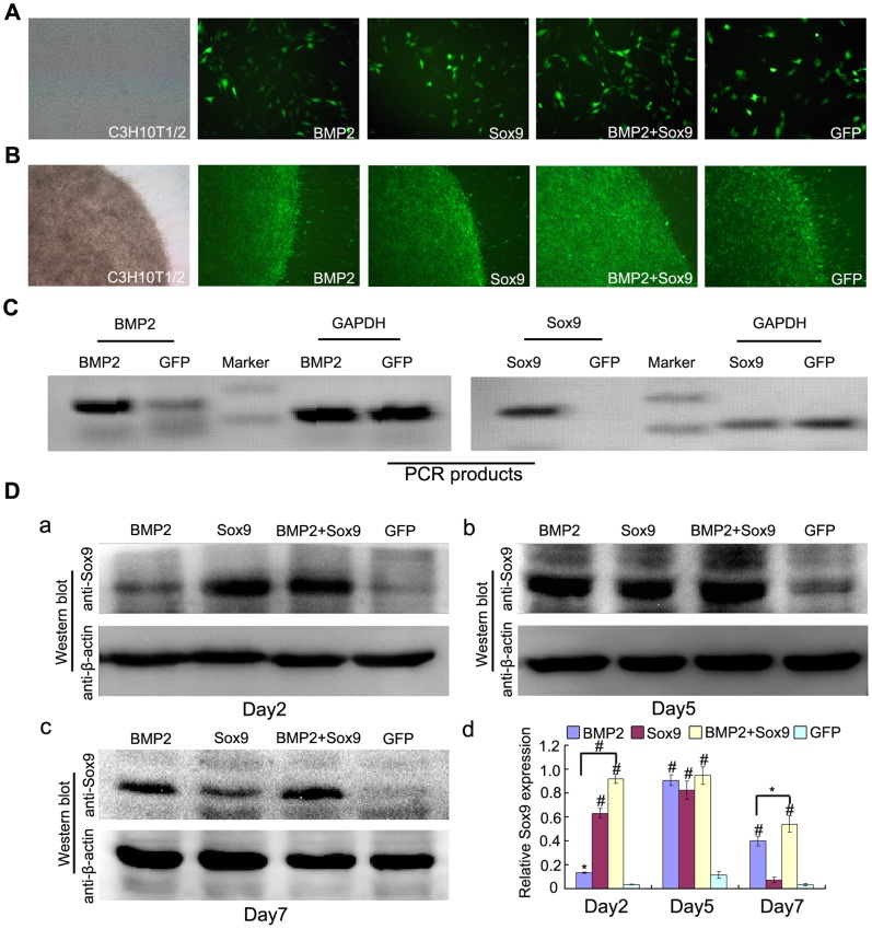
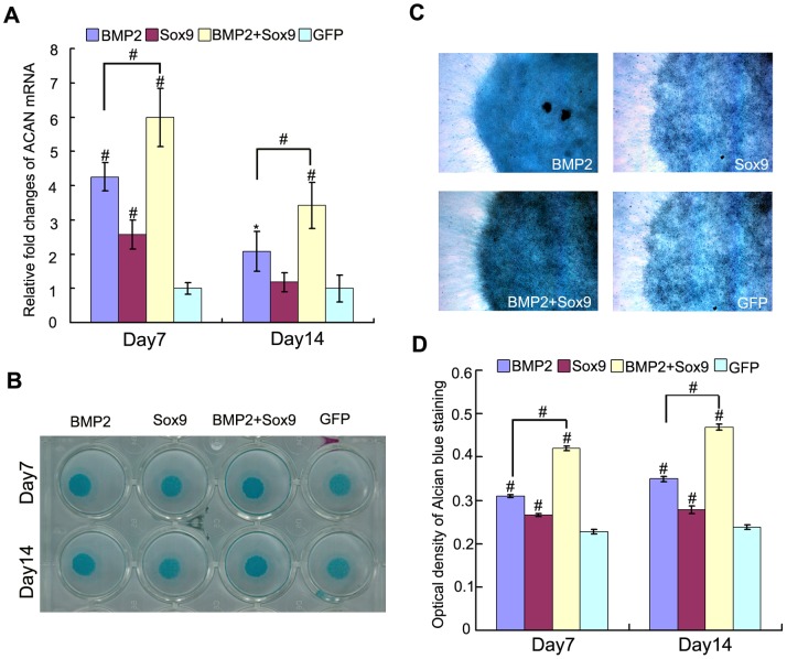
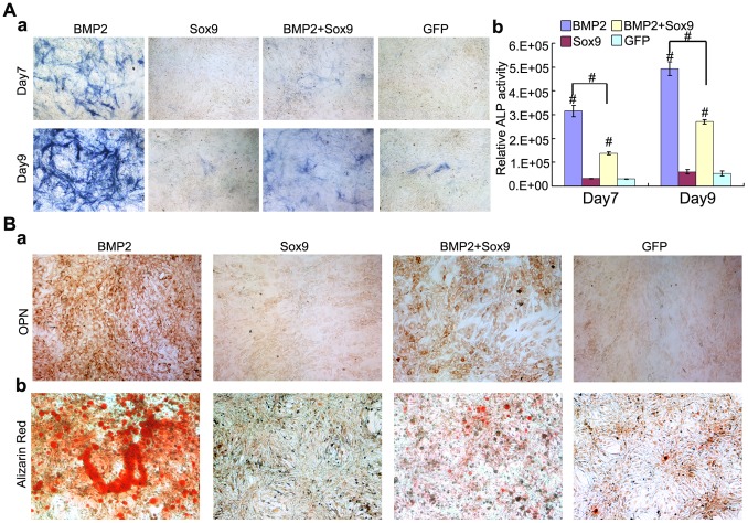
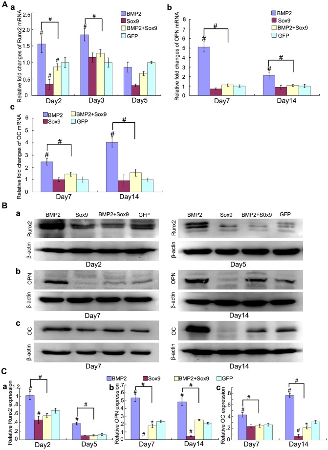
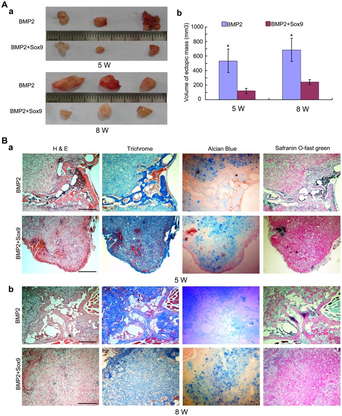
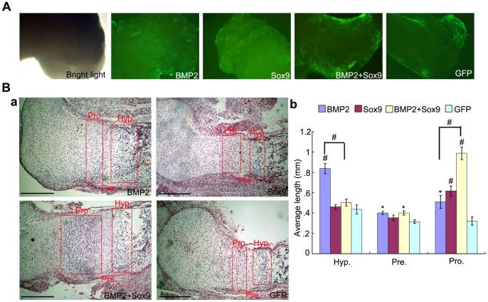
Bone morphogenetic protein 2 (BMP2) is one of the key chondrogenic growth factors involved in the cartilage regeneration. However, it also exhibits osteogenic abilities and triggers endochondral ossification. Effective chondrogenesis and inhibition of BMP2-induced osteogenesis and endochondral ossification can be achieved by directing the mesenchymal stem cells (MSCs) towards chondrocyte lineage with chodrogenic factors, such as Sox9. Here we investigated the effects of Sox9 on BMP2-induced chondrogenic and osteogenic differentiation of MSCs. We found exogenous overexpression of Sox9 enhanced the BMP2-induced chondrogenic differentiation of MSCs in vitro. Also, it inhibited early and late osteogenic differentiation of MSCs in vitro. Subcutaneous stem cell implantation demonstrated Sox9 potentiated BMP2-induced cartilage formation and inhibited endochondral ossification. Mouse limb cultures indicated that BMP2 and Sox9 acted synergistically to stimulate chondrocytes proliferation, and Sox9 inhibited BMP2-induced chondrocytes hypertrophy and ossification. This study strongly suggests that Sox9 potentiates BMP2-induced MSCs chondrogenic differentiation and cartilage formation, and inhibits BMP2-induced MSCs osteogenic differentiation and endochondral ossification. Thus, exogenous overexpression of Sox9 in BMP2-induced mesenchymal stem cells differentiation may be a new strategy for cartilage tissue engineering.


Conflict of interest statement

Competing Interests: The authors have declared that no competing interests exist.

Figures

Figure 1
Figure 1. Gene transduction and Sox9 expression in each treatment groups.
(A–B): Bright light and fluorescence microscope examination showed the transduction efficiency of recombinant adenoviruses in monolayer culture (24 hours after transduction, 100X) and micromass culture (3 days after transduction, 40X), respectively. (C): Recombinant adenoviruses mediated overexpression of BMP2 and Sox9 mRNA were evaluated by semi-quantitative RT-PCR analysis using GAPDH as a house keeping gene. (D): Sox9 expression were evaluated by western blot analysis in each treatment group at day 2, 5, and 7 after transduction (a to c) and relative Sox9 expression were analyzed by quantity one software using β-actin as controls (d), the results are expressed as mean±SD of triplicate experiments, *P<0.05, # P<0.01.
Figure 2
Figure 2. Sox9 potentiates BMP2-induced glycosaminoglycans synthesis in MSCs in micromass cultures.
(A): Real-time PCR for the expression of chondrogenic differentiation marker gene ACAN were conducted on day 7 and day 14 after infection of AdGFP, AdBMP2, and/or AdSox9, using GAPDH as a house keeping gene. (B–C): Alcian blue staining for sulfated glycosaminoglycans in micromass cultures of C3H10T1/2 cells on day 7 and day 14 after transduction of indicated recombinant adenoviruses, gross observation (B) and microscope examination (C, 40X) are shown. (D): Alcian blue staining quantifying: cells were extracted with 6 M guanidine hydrochloride, Optical density of the extracted dye was measured at 630 nm. The results were expressed as mean±SD of triplicate experiments, *P<0.05, # P<0.01.
Figure 3
Figure 3. Sox9 potentiates BMP2-induced Col2a1 synthesis in MSCs in micromass cultures.
(A): Real-time PCR for the expression of chondrogenic differentiation marker gene Col2a1 were conducted at continuously time points (from day 3 to day 14) after infection of AdGFP, AdBMP2, and/or AdSox9, using GAPDH as a house keeping gene. (B) Western blot for the expression of Col2a1 were conducted at continuously time points (from day 3 to day 14) after transduction of indicated recombinant adenoviruses (a), quantitatively, relative Sox9 expression were analyzed by quantity one software using β-actin as controls (b). The results are expressed as mean±SD of triplicate experiments, *P<0.05, # P<0.01.
Figure 4
Figure 4. Sox9 inhibits BMP2-induced early and late osteogenic differentiation of MSCs in vitro.
(A): C3H10T1/2 cells were infected with AdGFP, AdBMP2 and/or AdSox9. The ALP activities were measured on day 7 and day 9 using ALP histochemical staining (a), and chemiluminescent assays (b). (B): C3H10T1/2 cells were infected with indicated recombinant adenoviruses. On day 11 after infection, the expression of osteopontin (OPN) was assayed by immunocytochemical staining using anti-OPN antibody (a). For matrix mineralization, C3H10T1/2 cells were infected with indicated recombinant adenoviruses and cultured in mineralization medium. Alizarin Red staining were conducted on day 14 after infection (b). Each assay was done in triplicate. ALP assays results are expressed as mean±SD, *P<0.05, # P<0.01.
Figure 5
Figure 5. Sox9 inhibits BMP2-induced osteogenic markers expression in MSCs in vitro.
(A): Real-time PCR for the expression of early osteogenic differentiation gene Runx2 (Aa), late osteogenic gene osteocalcin (OC) and osteopontin (OPN) (b, c) were conducted at indicated time points after infection with AdGFP, AdBMP2, and/or AdSox9, using GAPDH as a house keeping gene. (B): Western blot for the expression of Runx2 (a), OPN (b) and OC (c) were conducted at indicated time points after transduction of indicated recombinant adenoviruses, respectively. (C) Relative protein expression was analyzed by quantity one software using β-actin as controls respectively. The results are expressed as mean±SD of triplicate experiments, *P<0.05, # P<0.01.
Figure 6
Figure 6. Sox9 potentiates BMP2-induced cartilage formation and inhibits BMP2-induced endochondral ossification in MSCs implantation in vivo.
(A): Macrographic images of ectopic masses. BMP2 or BMP2 and Sox9 co-infected C3H10T1/2 cells were implanted subcutaneously to the flanks of nude mice. Ectopic masses were retrieved at 5 weeks and 8 weeks (a). The volume of the masses was determined using vernier calipers (b). (B): Histological analysis of the retrieved samples. The retrieved samples were fixed, decalcified, paraffin-embedded and subjected to H&E, Masson’s Trichrome, Alcian blue and safranin O-fast green staining. Representative imagines are shown, magnification, 100X, scale bar = 1 mm.
Figure 7
Figure 7. Sox9 promotes expansion of the proliferating chondrocyte zone and inhibits BMP2-induced chondrocyte hypertrophy and ossification in organ cultures.
(A): Mouse E18.5 forelimbs (n = 4 each group) were harvested and transduced with AdGFP, AdBMP2, and/or AdSox9. The forelimbs were cultured in organ culture medium and the transduction efficiency was visualized under bright light and fluorescence microscope (40X). (B): Histological analysis of the cultured forelimbs. The forelimbs were fixed, decalcified, paraffin-embedded and subjected to H&E staining. Representative imagines are shown (a), magnification, 100X. The average length of the hypertrophic zones, prehypertropic zones and proliferating zones were also determined by using Image J software (b). Hyp = hypertrophic chondrocyte zone, Pre = pre-hypertrophic chondrocyte zone, Pro =  proliferating chondrocyte zone. *P<0.05, # P<0.01, scale bar = 1 mm.

Similar articles

Cited by

References

    1. Steinert AF, Noth U, Tuan RS (2008) Concepts in gene therapy for cartilage repair. Injury 39 Suppl 1: S97–113. - PMC - PubMed
    1. Benthien JP, Schwaninger M, Behrens P (2011) We do not have evidence based methods for the treatment of cartilage defects in the knee. Knee surgery, sports traumatology, arthroscopy : official journal of the ESSKA 19: 543–552. - PubMed
    1. Rodriguez-Merchan EC (2012) Regeneration of articular cartilage of the knee. Rheumatology international. - PubMed
    1. Kao YJ, Ho J, Allen CR (2011) Evaluation and management of osteochondral lesions of the knee. The Physician and sportsmedicine 39: 60–69. - PubMed
    1. Chen H, Sun J, Hoemann CD, Lascau-Coman V, Ouyang W, et al. (2009) Drilling and microfracture lead to different bone structure and necrosis during bone-marrow stimulation for cartilage repair. Journal of orthopaedic research : official publication of the Orthopaedic Research Society 27: 1432–1438. - PubMed

Publication types

MeSH terms