Clinical Trial
. 2014 Feb 13;8(2):e2694.
doi: 10.1371/journal.pntd.0002694. eCollection 2014 Feb.

Rapid healing of cutaneous leishmaniasis by high-frequency electrocauterization and hydrogel wound care with or without DAC N-055: a randomized controlled phase IIa trial in Kabul

Affiliations
Clinical Trial

Rapid healing of cutaneous leishmaniasis by high-frequency electrocauterization and hydrogel wound care with or without DAC N-055: a randomized controlled phase IIa trial in Kabul

Ahmad Fawad Jebran et al. PLoS Negl Trop Dis. .

Abstract

Background: Anthroponotic cutaneous leishmaniasis (CL) due to Leishmania (L.) tropica infection is a chronic, frequently disfiguring skin disease with limited therapeutic options. In endemic countries healing of ulcerative lesions is often delayed by bacterial and/or fungal infections. Here, we studied a novel therapeutic concept to prevent superinfections, accelerate wound closure, and improve the cosmetic outcome of ACL.

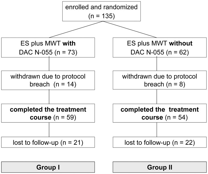
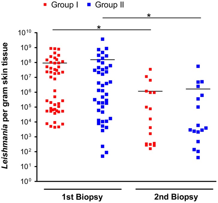
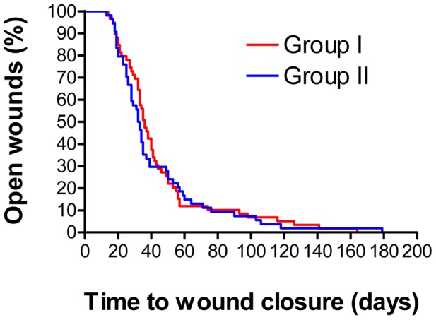
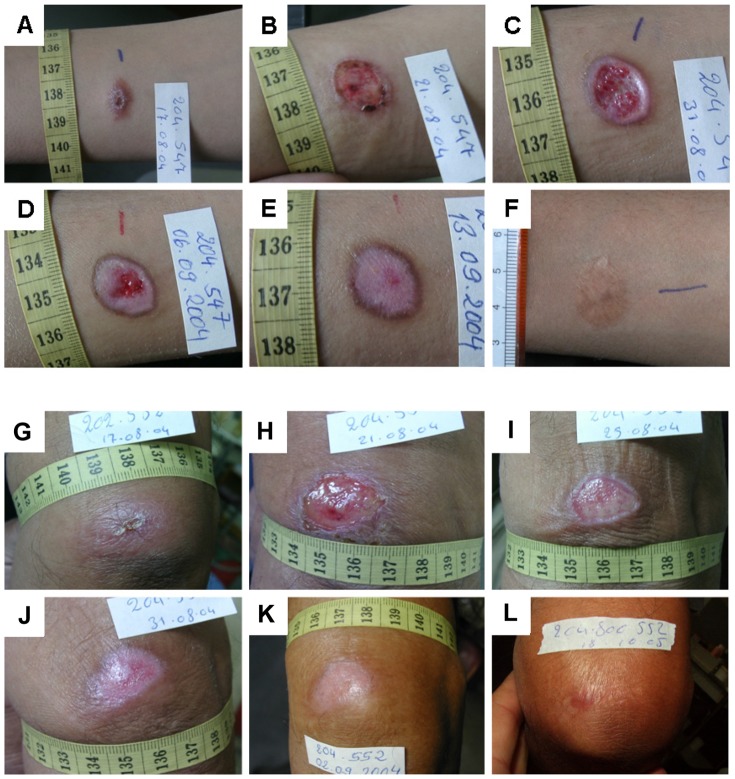
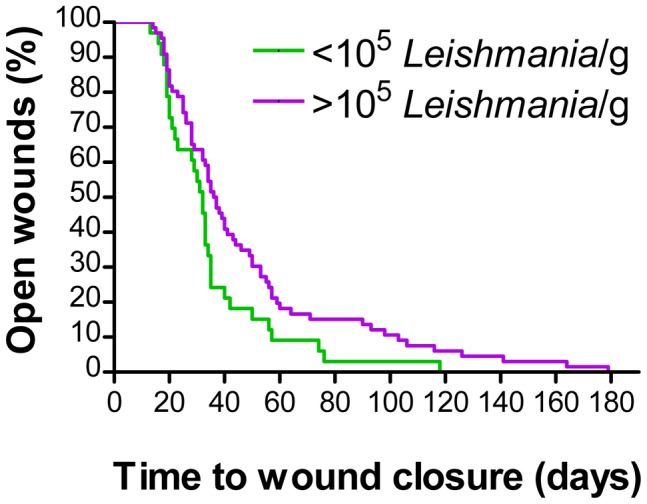
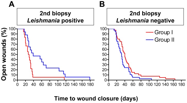
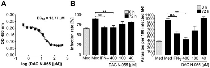
Methodology/principal findings: From 2004 to 2008 we performed a two-armed, randomized, double-blinded, phase IIa trial in Kabul, Afghanistan, with patients suffering from L. tropica CL. The skin lesions were treated with bipolar high-frequency electrocauterization (EC) followed by daily moist-wound-treatment (MWT) with polyacrylate hydrogel with (group I) or without (group II) pharmaceutical sodium chlorite (DAC N-055). Patients below age 5, with facial lesions, pregnancy, or serious comorbidities were excluded. The primary, photodocumented outcome was the time needed for complete lesion epithelialization. Biopsies for parasitological and (immuno)histopathological analyses were taken prior to EC (1(st)), after wound closure (2(nd)) and after 6 months (3(rd)). The mean duration for complete wound closure was short and indifferent in group I (59 patients, 43.1 d) and II (54 patients, 42 d; p = 0.83). In patients with Leishmania-positive 2(nd) biopsies DAC N-055 caused a more rapid wound epithelialization (37.2 d vs. 58.3 d; p = 0.08). Superinfections occurred in both groups at the same rate (8.8%). Except for one patient, reulcerations (10.2% in group I, 18.5% in group II; p = 0.158) were confined to cases with persistent high parasite loads after healing. In vitro, DAC N-055 showed a leishmanicidal effect on pro- and amastigotes.

Conclusions/significance: Compared to previous results with intralesional antimony injections, the EC plus MWT protocol led to more rapid wound closure. The tentatively lower rate of relapses and the acceleration of wound closure in a subgroup of patients with parasite persistence warrant future studies on the activity of DAC N-055.

Trial registration: ClinicalTrials.gov NCT00947362.


Conflict of interest statement

The authors have declared that no competing interests exist.

Figures

Figure 1
Figure 1. Study flow diagram showing the enrolment, randomization and follow-up of patients (CONSORT flow chart).
Figure 2
Figure 2. Parasite burden of the lesions before treatment (1st biopsy) and after wound closure (2nd biopsy).
▪ Patients, who received EC plus MWT with DAC N-055 (group I); ▪ patients, who received EC plus MWT without DAC N-055 (group II). Each symbol (▪, ▪) represents one biopsy; horizontal lines mark the mean value of each group. After wound closure (2nd biopsy) 42 out of 59 biopsies in group I and 37 out of 54 in group II were Leishmania-negative (not shown). Level of significance was estimated using Student's t-test (*, p<0.05).
Figure 3
Figure 3. Wound healing in patients who received EC/MWT with (group I) or without DAC N-055 (group II).
The data are presented in the format of a Kaplan-Meier analysis. No statistical difference was found in the log rank test Mantel Cox (p = 0.638).
Figure 4
Figure 4. Representative examples of the tissue repair process and wound healing in the two treatment groups.
A–F: 13 years old male CL patient treated with EC/MWT with DAC N-055. A: Ulcerated lesion before treatment located on the forearm. B: 4 d after EC, C: Formation of granulation tissue and beginning epithelialization after 15 d, D: Progressing epithelialization (d 19), E: Complete wound closure after 27 d, F: Follow-up after 20 months revealed a flat scar with hyperpigmentation. G–L: 63 years old male CL patient treated with EC/MWT without DAC N-055. G: Lesion on the upper arm with early ulceration prior to treatment. H: 4 d after EC, I: Formation of granulation tissue covered with fibrin and beginning epithelialization after 8 d, J: Progressing epithelialization (14 d), K: Complete wound closure after 16 d, L: Follow-up after 14 months revealed a flat scar.
Figure 5
Figure 5. Impact of the lesional parasite load prior to treatment on the wound healing.
The wound healing for patients of both treatment arms (PP analysis) with a parasite load higher of lower than 105 per gram lesion tissue (1st biopsy) was analysed using Kaplan-Meier curves. The log rank test Mantel-Cox revealed a significant difference (p<0.05).
Figure 6
Figure 6. Kaplan-Meier analysis of the wound healing depending on the parasite status after treatment.
The patients of both treatment arms (PP analysis) were grouped with respect to the presence or absence of Leishmania parasites in the 2nd biopsy (i.e. after treatment). The wound healing was analysed using Kaplan-Meier curves. A: 2nd biopsy Leishmania-positive (p = 0.08; log rank test Mantel-Cox). B: 2nd biopsy Leishmania-negative (p = 0.2; log rank test Mantel-Cox).
Figure 7
Figure 7. Effect of DAC N-055 on L. tropica parasites.
A: Extracellular L. tropica promastigotes. The survival of extracellular Leishmania promastigotes after 4 d of incubation with DAC N-055 was determined via optical density at 450 nm. The OD values were logarithmically transformed and a curve fit was carried out. The mean of the curve (solid line) and the 95% confidence interval (dashed line) of two independent experiments with 24 OD values per DAC N-055 concentration as well as the ED50 are given. B: Intracellular L. tropica amastigotes. After infection of adherent human monocytes (macrophages, MΦ) for 19 h with stationary phase-grown and opsonized L. tropica promastigotes (MOI = 5) the cells were washed and stimulated for 72 h as indicated. The time point directly after the pulse infection was defined as 0 h. The infection rate and the number of parasites per 100 infected MΦ were determined at time point 0 h and after 72 h of stimulation. The mean ± SEM of three independent experiments are shown. For statistical analysis the Mann-Whitney test was used (** = p<0.001, n. s. = not significant).

References

    1. Bailey MS, Lockwood DN (2007) Cutaneous leishmaniasis. Clin Dermatol 25: 203–211. - PubMed
    1. Reithinger R, Dujardin JC, Louzir H, Pirmez C, Alexander B, et al. (2007) Cutaneous leishmaniasis. Lancet Infect Dis 7: 581–596. - PubMed
    1. WHO (2010) Control of Leishmaniasis. WHO Technical Report Series 949: 1–187.
    1. Bogdan C (2012) Leishmaniasis in Rheumatology, Hematology, and Oncology: Epidemiological, Immunological, and Clinical Aspects and Caveats. Ann Rheumatic Diseases 71 (suppl. 2) i60–i66. - PubMed
    1. Kibbi AG, Karam PG, Kurban AK (1987) Sporotrichoid leishmaniasis in patients from Saudi Arabia: clinical and histologic features. J Am Acad Dermatol 17: 759–764. - PubMed

Publication types

Associated data