. 2014 Jan 15;7(2):537-51.
eCollection 2014.

NOX1 is responsible for cell death through STAT3 activation in hyperoxia and is associated with the pathogenesis of acute respiratory distress syndrome

Affiliations

NOX1 is responsible for cell death through STAT3 activation in hyperoxia and is associated with the pathogenesis of acute respiratory distress syndrome

Stephanie Carnesecchi et al. Int J Clin Exp Pathol. .

Abstract

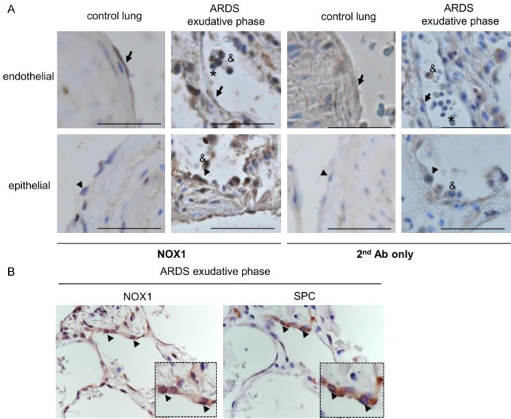
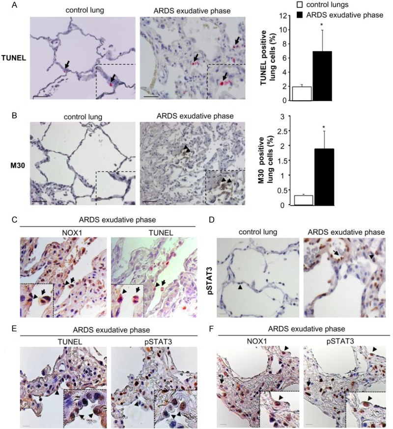
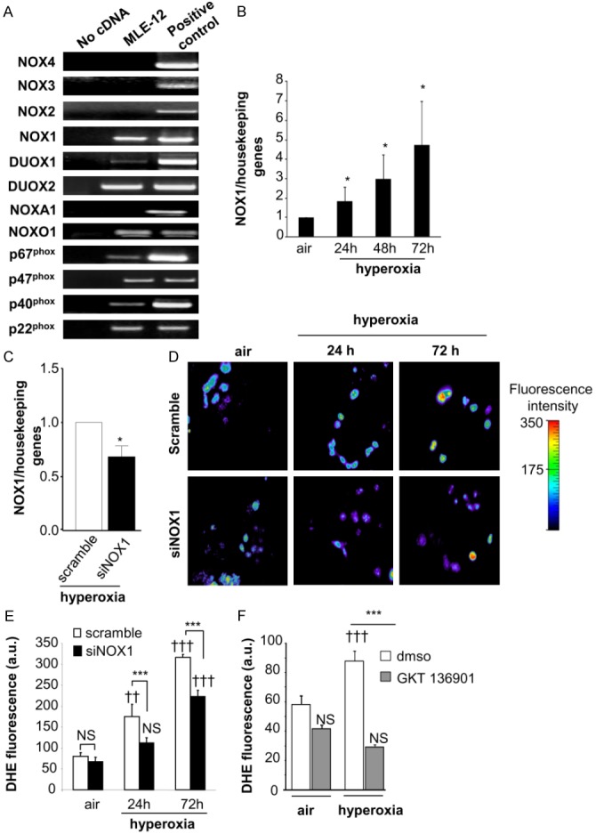
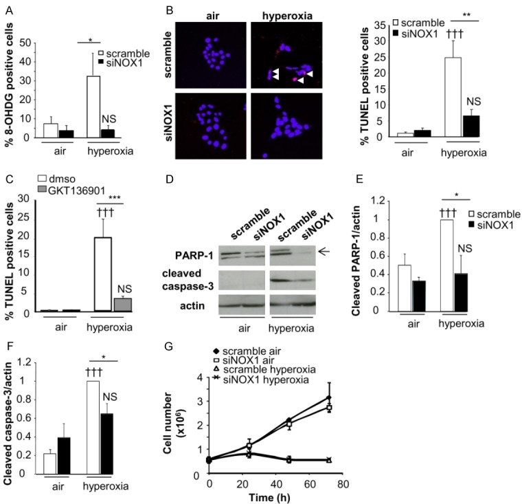
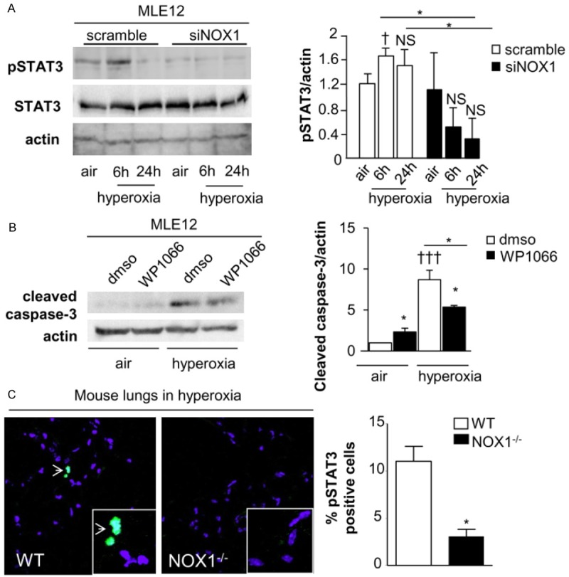
Reactive oxygen species (ROS) contribute to alveolar cell death in acute respiratory distress syndrome (ARDS) and we previously demonstrated that NOX1-derived ROS contributed to hyperoxia-induced alveolar cell death in mice. The study investigates whether NOX1 expression is modulated in epithelial cells concomitantly to cell death and associated to STAT3 signaling in the exudative phase of ARDS. In addition, the role of STAT3 activation in NOX1-dependent epithelial cell death was confirmed by using a lung epithelial cell line and in mice exposed to hyperoxia. NOX1 expression, cell death and STAT3 staining were evaluated in the lungs of control and ARDS patients by immunohistochemistry. In parallel, a stable NOX1-silenced murine epithelial cell line (MLE12) and NOX1-deficient mice were used to characterize signalling pathways. In the present study, we show that NOX1 is detected in alveolar epithelial cells of ARDS patients in the exudative stage. In addition, increased alveolar epithelial cell death and phosphorylated STAT3 are observed in ARDS patients and associated with NOX1 expression. Phosphorylated STAT3 is also correlated with TUNEL staining. We also confirmed that NOX1-dependent STAT3 activation participates to alveolar epithelial cell death. Silencing and acute inhibition of NOX1 in MLE12 led to decreased cell death and cleaved-caspase 3 induced by hyperoxia. Additionally, hyperoxia-induced STAT3 phosphorylation is dependent on NOX1 expression and associated with cell death in MLE12 and mice. This study demonstrates that NOX1 is involved in human ARDS pathophysiology and is responsible for the damage occurring in alveolar epithelial cells at least in part via STAT3 signalling pathways.

Keywords: ARDS; NOX1; ROS; STAT3; cell death; hyperoxia.


Figures

Figure 1
Figure 1
NOX1 is highly expressed in alveolar epithelium of ARDS patients. ARDS lung tissues were analyzed for NOX1 expression by immunohistochemistry. (A) Lung structures from control and ARDS in exudative phase were stained with anti-NOX1 antibody (NOX1 Ab) or with secondary antibody only (2nd Ab only). In control lungs, the anti-NOX1 antibody stained pulmonary endothelial cells (arrow) but not epithelial cells (type II, arrowhead). In the exudative stage of ARDS, both epithelial cells (type II, arrowhead) and endothelial cells (arrow) were positive. Note the presence of NOX1 in macrophages (ampersand), but not in neutrophils (asterisk) located inside the alveoli of ARDS lungs. Scale bars, 100 μm. (B) Serial lung sections of ARDS in the exudative phase were stained with NOX1 antibody and pro-surfactant C (a specific marker of Type II epithelial cells). Epithelial type II cells are positive for NOX1 in the exudative stage of ARDS (arrowhead, two different magnifications), Scale bars, 50 μm.
Figure 2
Figure 2
Epithelial cell death detected in early phase of ARDS is associated with NOX1 and pSTAT3. Cell death was analyzed in control and ARDS lungs during exudative phases by TUNEL and M30 staining. (A) Representative images of control and ARDS lungs sections stained with TUNEL and (B) M30. TUNEL-positive cells appear in pink (arrow) and M30-positive cells are in brown (arrowhead). Scale bars, 50 μm. The numbers of TUNEL- and M30-positive cells are expressed as percent of all nuclei counted in lung sections. Quantification of positive staining was performed using Metamorph analysis software (10 images per subjects, 3-4 subjects per group) P=NS, *P<0.05, ARDS versus control patients. (C) Immunostaining for NOX1 (brown) and TUNEL (pink) were done on serial lung sections of ARDS patients in the exudative phase (two different magnifications). Note the presence of NOX1 in the TUNEL-positive epithelial cells (arrowhead) and macrophages (arrow). Scale bars, 50 μm. (D) Control and ARDS lung tissues were analyzed for phosphorylated STAT3 by immunohistochemistry. In control lungs, STAT3 phosphorylation was not detected in epithelial (arrowhead) and endothelial (data not shown) cells whereas in ARDS lung sections, epithelial (arrowhead) and endothelial (arrow) cells were positive for phosphorylated STAT3 in the exudative phase. (E) Serial lung sections of ARDS exudative phase were stained with an anti-phosphorylated STAT3 antibody and TUNEL, or (F) with an anti-NOX1 antibody. Phosphorylated STAT3-positive cells were also positive for both TUNEL and NOX1 (arrowheads and arrow), Scale bars, 50 μm.
Figure 3
Figure 3
Hyperoxia increases NOX1 mRNA expression and ROS-dependent NOX1 production in MLE12. (A) Expression of NOX isoforms in MLE12. NOX1, DUOX1/2, and the regulatory subunits p22phox, p40phox, p67phox and NOXO1 were detected in MLE12 by RT-PCR. Lung tissues were used as positive control for the detection of NOX1, 2, 4, DUOX1/2, and the regulatory subunits. For the detection of NOX3 mRNA expression, ear tissue was used as positive control. Absence of cDNA was used as negative control for expression of NOX1 mRNA in MLE12 measured by real time PCR in air condition and (B) at 24, 48 and 72 h following hyperoxia exposure (n=3). *P<0.05. (C) NOX1 mRNA measured by qualitative RT-PCR in scramble- and NOX1-silenced MLE12 (siNOX1) at 72 h of hyperoxia. (D, E) Representative fluorescent images of scramble- and NOX1-silenced MLE12 loaded with DHE. ROS production was measured by analysing DHE staining (10 M) in scramble- and NOX1-silenced MLE12 in air or hyperoxia for 24 and 72 h, and visualized by confocal microscopy (pseudocolor). Original magnification, X40. Fluorescence intensity was quantified in MLE12, bars represent the mean ± SEM (n>50 cells for each group; P=NS, ***P<0.001, scramble-versus NOX1-silenced cells under hyperoxia; ††P<0.01, †††P<0.001, air versus hyperoxia). (F) Acute inhibition of NOX1 prevents hyperoxia-induced ROS production. MLE12 were treated with dmso, or GKT136901 (10 μm) 1 hour before hyperoxia exposure and for 72 h. ROS generation was measured by analysing DHE fluorescence intensity in treated MLE12 exposed to air or hyperoxia for 72 h. Bars represent the mean ± SEM (n>50 cells for each group; ***P<0.001 cells treated with NOX inhibitor compared to cells treated with DMSO exposed to hyperoxia; P=NS, †††P<0.001, air versus hyperoxia).
Figure 4
Figure 4
Acute and stable NOX1 inhibition decrease hyperoxia-induced death of MLE12. Cell death was evaluated in control and NOX1-silenced MLE12 in air or hyperoxic condition. A: Transduced MLE12 exposed to air or hyperoxia for 72 h were stained with 8-hydroxy-2’-deoxyguanosine antibody (8-OHdG, red) and DAPI (blue) and the number of 8-OHdG-positive cells is expressed as percent of all nuclei (n>50 for each group, 3 independent experiments). B: Representative images of transduced MLE12 stained with TUNEL (red) and DAPI (blue) at 72 h of air or hyperoxia. White arrows indicate TUNEL-positive cells which appear in pink. The number of TUNEL-positive cells is expressed as percent of all nuclei (n>50 for each group, 3 independent experiments). P=NS, †††P<0.001 air versus hyperoxia; ***P<0.001, **P<0.01, *P<0.05 scramble-versus NOX1-silenced cells in hyperoxia. C: MLE12 were treated with DMSO, or GKT136901 (10 m) and exposed to hyperoxia for 72 h. ***P<0.001 cells treated with NOX inhibitor compared to cells exposed to DMSO in hyperoxia. P=NS, †††P<0.001 air versus hyperoxia. D-F: Protein lysate of transduced MLE12 were blotted for cleaved caspase-3 and PARP-1 and quantified by densitometry (right panel; n=3). β-actin was used to control equal loading. P=NS, †††P<0.001 air versus hyperoxia; *P<0.05 scramble-versus NOX1-silenced cells in hyperoxia. G: Cell growth was measured by using sulforhodamine B for different times. Absorbance was measured at 490 nm and cell number was determined. The relationship between cell number (protein content per well) and absorbance is linear from 0 to 5.106 cells. No difference was observed between scramble-versus NOX1-silenced cells in hyperoxia.
Figure 5
Figure 5
STAT3 phosphorylation is dependent on NOX1 and participates to cell death in hyperoxic condition. A: Western-blot of phosphorylated and total STAT3 in MLE12 were quantified by densitometry (n=3). β-actin was used to control equal loading. Phosphorylated forms of the respective proteins are indicated by the prefix “p”. P=NS, P=NS, †P<0.05 air versus hyperoxia *P<0.05 scramble-versus NOX1-silenced cells in hyperoxia at different time points. B: MLE12 was pre-treated with DMSO or WP1066 (1 m) 6 hours before hyperoxia and for 72 h. Western-blot of cleaved-caspase 3 was quantified by densitometry (n=3). β-actin was used to control equal loading †††P<0.001 cells treated with STAT3 inhibitor compared to cells exposed to DMSO in hyperoxia, *P<0.05 cells exposed to DMSO compared to cells treated to STAT3 inhibitor in air, and cells treated to STAT3 inhibitor in air compared to cells treated with STAT3 inhibitor in hyperoxia. C: Representative images of mouse lung sections from WT and NOX1-deficient mice stained with anti-pSTAT3 (green) and DAPI (blue). Original magnification, X 40. The number of pSTAT3-positive cells is expressed as percent of all nuclei (n=3 mice), *P<0.05 WT-versus NOX1-deficient mice exposed to hyperoxia for 72 h.

References

    1. Ware LB, Matthay MA. The acute respiratory distress syndrome. N Engl J Med. 2000;342:1334–1349. - PubMed
    1. Kallet RH, Matthay MA. Hyperoxic acute lung injury. Respir Care. 2013;58:123–141. - PMC - PubMed
    1. Lee KS, Choi YH, Kim YS, Baik SH, Oh YJ, Sheen SS, Park JH, Hwang SC, Park KJ. Evaluation of bronchoalveolar lavage fluid from ARDS patients with regard to apoptosis. Respir Med. 2008;102:464–469. - PubMed
    1. Lamb NJ, Gutteridge JM, Baker C, Evans TW, Quinlan GJ. Oxidative damage to proteins of bronchoalveolar lavage fluid in patients with acute respiratory distress syndrome: evidence for neutrophil-mediated hydroxylation, nitration, and chlorination. Crit Care Med. 1999;27:1738–1744. - PubMed
    1. Carnesecchi S, Pache JC, Barazzone-Argiroffo C. NOX enzymes: potential target for the treatment of acute lung injury. Cell Mol Life Sci. 2012;69:2373–2385. - PMC - PubMed

Publication types

MeSH terms

LinkOut - more resources