. 2014 Jan 15;7(2):552-64.
eCollection 2014.

Alveolar epithelial cells in idiopathic pulmonary fibrosis display upregulation of TRAIL, DR4 and DR5 expression with simultaneous preferential over-expression of pro-apoptotic marker p53

Affiliations

Alveolar epithelial cells in idiopathic pulmonary fibrosis display upregulation of TRAIL, DR4 and DR5 expression with simultaneous preferential over-expression of pro-apoptotic marker p53

Khondoker M Akram et al. Int J Clin Exp Pathol. .

Abstract

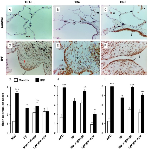
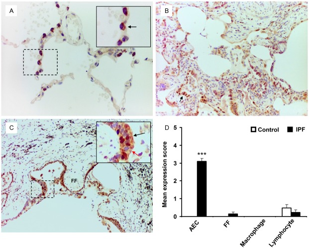
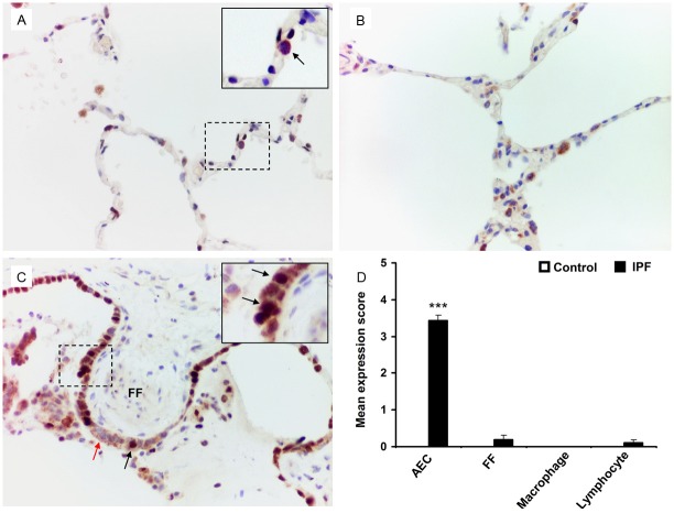
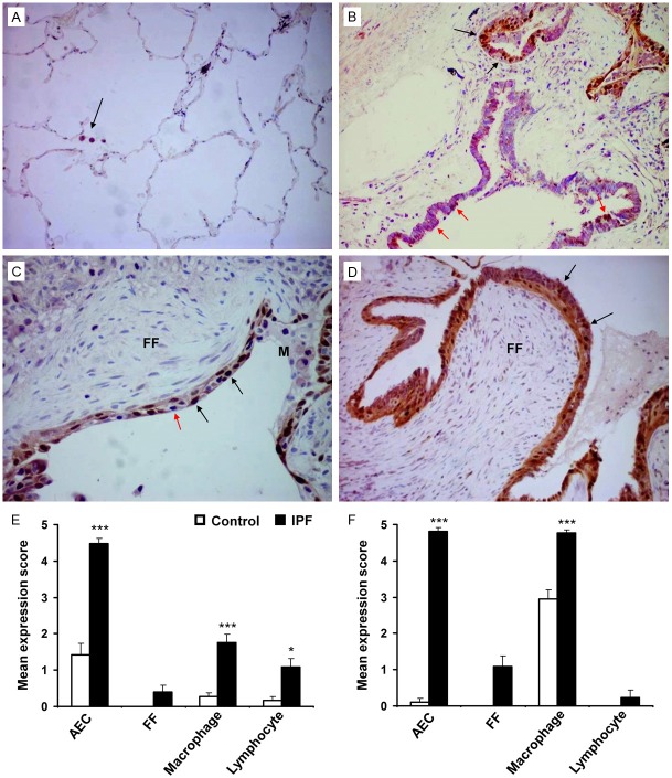
Idiopathic pulmonary fibrosis (IPF) is a progressive, debilitating, and fatal lung disease of unknown aetiology with no current cure. The pathogenesis of IPF remains unclear but repeated alveolar epithelial cell (AEC) injuries and subsequent apoptosis are believed to be among the initiating/ongoing triggers. However, the precise mechanism of apoptotic induction is hitherto elusive. In this study, we investigated expression of a panel of pro-apoptotic and cell cycle regulatory proteins in 21 IPF and 19 control lung tissue samples. We reveal significant upregulation of the apoptosis-inducing ligand TRAIL and its cognate receptors DR4 and DR5 in AEC within active lesions of IPF lungs. This upregulation was accompanied by pro-apoptotic protein p53 overexpression. In contrast, myofibroblasts within the fibroblastic foci of IPF lungs exhibited high TRAIL, DR4 and DR5 expression but negligible p53 expression. Similarly, p53 expression was absent or negligible in IPF and control alveolar macrophages and lymphocytes. No significant differences in TRAIL expression were noted in these cell types between IPF and control lungs. However, DR4 and DR5 upregulation was detected in IPF alveolar macrophages and lymphocytes. The marker of cellular senescence p21(WAF1) was upregulated within affected AEC in IPF lungs. Cell cycle regulatory proteins Cyclin D1 and SOCS3 were significantly enhanced in AEC within the remodelled fibrotic areas of IPF lungs but expression was negligible in myofibroblasts. Taken together these findings suggest that, within the remodelled fibrotic areas of IPF, AEC can display markers associated with proliferation, senescence, and apoptotosis, where TRAIL could drive the apoptotic response. Clear understanding of disease processes and identification of therapeutic targets will direct us to develop effective therapies for IPF.

Keywords: DR4; DR5; Idiopathic pulmonary fibrosis; TRAIL; immunohistochemistry; p21WAF1; p53.


Figures

Figure 1
Figure 1
Immunohistochemical analysis of IPF and control lungs for expression of TRAIL and its receptors DR4 and DR5. (A, D) TRAIL expression detection in the alveolar regions of normal (A) and IPF lung tissue (D). Negligible TRAIL expression was detected in control lung but was highly expressed in AEC (black arrow) and myofibroblasts (red arrow) of IPF lung. (B, E) Immunostaing for DR4 on normal (B) and IPF (E) lung tissue. High level of DR4 expression was detected in AEC over the fibrotic foci (FF), myofibroblasts and macrophages (M) of IPF lung (E), arrow indicates DR4 positive AEC. (C, F) Immunostaing for DR5 on normal (C) and IPF (F) lung tissue. Bronchiolar epithelial cells stains positive for DR5 (arrow) but AEC are negative in control lung. DR5 positively stained macrophages (M) are observed in control lung. Significantly increased DR5 expression is detected in AEC (arrows), myofibroblasts, macrophages and lymphocytes (F). (G-I) Mean expression score profiles for TRAIL (G), DR4 (H) and DR5 (I) on control (open bar) and IPF (black bar) lungs respectively. FF indicates myofibroblasts within the fibroblastic foci. Image magnification 400x (A-C) and 200x (D-F). *p<0.05, **p<0.001, ***p<0.0001 vs control. ns = not significant, M = macrophages.
Figure 2
Figure 2
Dual-labelled immunohistochemistry of IPF and control lung samples for p53 and SP-C expression. (A) SP-C cytoplasmic stained (brown) AEC are negative for p53 nuclear staining (arrow, negative blue nucleus). (B) Patchy nuclear expression of p53 (very intense purple) is observed in SP-C positive AEC within the fibrotic lesions of IPF lung samples (arrow). (C) Increased level of dual-positive p53/SP-C AEC are detected at the areas covering fibroblastic foci (FF) (inset, black arrow indicates dual positive cells, red arrow indicates p53 negative but SP-C positive AEC). Myofibroblasts within the fibroblastic foci (FF) are negative for both p53 and SP-C. (D) Mean expression score profiles for p53 on control (open bar) and IPF (black bar) lung tissue samples respectively. FF indicates myofibroblasts within the fibroblastic foci. Image magnification 400x (A), 200x (B, C). ***p<0.0001 vs control.
Figure 3
Figure 3
Dual TUNEL and immunohistochemistry with proSP-C on human lung tissue samples. TUNEL positive AEC were not detected in control lung tissues (A). Increased number of TUNEL positive (green nuclei, arrows) AEC (red cytoplasmic stain) were detected in IPF lung tissue sample (B).
Figure 4
Figure 4
Dual-labelled immunohistochemistry of IPF and control lung samples for p21WAF1 and SP-C expression. A. SP-C cytoplasmic stained (brown) AEC show negative staining for p21WAF1 nuclear staining (arrow) in control lung. B. A representative image of conserved area of IPF lung demonstrating no p21WAF1 expression. C. Increased level of dual-positive p21WAF1/SP-C AEC (VIP stain showing dark brown against brown cytoplasmic background) are detected at the areas covering fibroblastic foci (FF) (black arrows). p21WAF1 negative but SP-C positive type II AEC are shown by red arrow. D. Mean expression score profiles for p21WAF1 on control (open bar) and IPF (black bar) lung tissue samples respectively. FF indicates myofibroblasts within the fibroblastic foci. Image magnification 400x for all. ***p<0.0001 vs control.
Figure 5
Figure 5
Dual-labelled immunohistochemistry of IPF and control lung samples for Cyclin D1 and SOCS3 expression. (A) Representative histological image of control lung tissue shows no Cyclin D1 or SOCS expression within the alveoli. SOCS3 expression is expressed by macrophages (purple cytoplasmic stain) (arrow). (B) IPF lung tissue demonstrates dual expression of nuclear Cyclin D1 (brown) and cytoplasmic SOCS3 (purple) in the hyperplastic AEC (black arrows). Extensive expression of SOCS3 within the ciliated epithelium was also noted (red arrows). (C, D) Two separate IPF tissue samples with AEC overlying fibroblastic foci expressing both Cyclin D1 (brown) and SOCS3 (purple), however, cells within the foci did not express either marker. Red arrow indicates dual-negative AEC (C). (E, F) Mean expression score profiles for Cyclin D1 and SOCS3 on 19 control (open bar) and 21 IPF (black bar) lung tissue samples respectively. FF indicates myofibroblasts within the fibroblastic foci. Image magnification 200x (A), 400x (B-D). *p<0.05, ***p<0.0001 vs control. M = macrophages.

Similar articles

Cited by

References

    1. Raghu G, Collard HR, Egan JJ, Martinez FJ, Behr J, Brown KK, Colby TV, Cordier JF, Flaherty KR, Lasky JA, Lynch DA, Ryu JH, Swigris JJ, Wells AU, Ancochea J, Bouros D, Carvalho C, Costabel U, Ebina M, Hansell DM, Johkoh T, Kim DS, King TE Jr, Kondoh Y, Myers J, Müller NL, Nicholson AG, Richeldi L, Selman M, Dudden RF, Griss BS, Protzko SL, Schünemann HJ ATS/ERS/JRS/ALAT Committee on Idiopathic Pulmonary Fibrosis. ATS/ERS/JRS/ALAT statement: idiopathic pulmonary fibrosis: evidence-based guidelines for diagnosis and management. Am J Respir Crit Care Med. 2011;183:788–824. - PMC - PubMed
    1. Gribben J, Hubbard RB, Le-Jeune I, Smith CJ, West J, Tata LJ. Incidence and mortality of idiopathic pulmonary fibrosis and sarcoidosis in the UK. Thorax. 2006;61:980–985. - PMC - PubMed
    1. Dacic S, Yousem SA. Histologic classification of idiopathic chronic interstitial pneumonias. Am J Respir Cell Mol Biol. 2003;29:S5–S9. - PubMed
    1. Maher TM, Wells AU, Laurent GJ. Idiopathic pulmonary fibrosis: multiple causes and mechanisms? Eur Respir J. 2007;30:835–839. - PubMed
    1. Selman M, King TE, Pardo A. American Thoracic Society, European Respiratory Society & American College of Chest Physicians. Idiopathic pulmonary fibrosis: prevailing and evolving hypotheses about its pathogenesis and implications for therapy. Ann Intern Med. 2001;134:136–151. - PubMed

Publication types

MeSH terms

Substances

LinkOut - more resources