

microRNA-21 promotes cardiac fibrosis and development of heart failure with preserved left ventricular ejection fraction by up-regulating Bcl-2
- PMID: 24551276
- PMCID: PMC3925900
microRNA-21 promotes cardiac fibrosis and development of heart failure with preserved left ventricular ejection fraction by up-regulating Bcl-2
Abstract
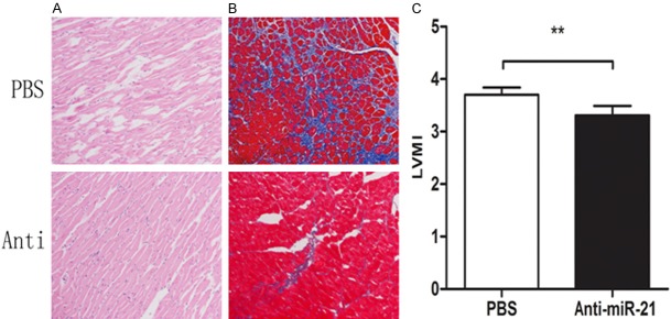
The morbidity and mortality of heart failure with preserved left ventricular ejection fraction (HFpEF) were similar to those of systolic heart failure, but the pathogenesis of HFpEF remains poorly understood. It was demonstrated that, in systolic heart failure, microRNA-21 (miR-21) could inhibit the apoptosis of cardiac fibroblasts, leading to cardiac hypertrophy and myocardial fibrosis, but the role of miR-21 in HFpEF remains unknown. By employing cell culture technique, rat myocardiocytes and cardiac fibroblasts were obtained. The expression of miR-21 in the two cell types under different conditions was compared and we found that the miR-21 expression was significantly higher in cardiac fibroblasts than in myocardiocytes. We established a rat HFpEF model and harvested the tissues of cardiac apex for pathological examination, Northern blotting and so forth. We found that miR-21 expression was significantly higher in model rats than in sham-operated rats, and the model rats developed the cardiac atrophy and cardiac fibrosis. After injection of miR-21 antagonist, the the cardiac atrophy and cardiac fibrosis were conspicuously ameliorated. Both in vivo and in vitro, inhibition of miR-21 expression resulted in reduced Bcl-2 expression while over-expression of miR-21 led to elevation of Bcl-2 expression. Our study suggested that miR-21 promoted the development of HFpEF by up-regulating the expression of anti-apoptotic gene Bcl-2 and thereby suppressing the apoptosis of cardiac fibrosis.
Keywords: Bcl-2; Cardiac fibrosis; HFpEF; miR-21.
Figures


Similar articles
-
Ivabradine Ameliorates Cardiac Function in Heart Failure with Preserved and Reduced Ejection Fraction via Upregulation of miR-133a.Oxid Med Cell Longev. 2021 Sep 29;2021:1257283. doi: 10.1155/2021/1257283. eCollection 2021. Oxid Med Cell Longev. 2021. PMID: 34630844 Free PMC article.
-
Prevention of heart failure with preserved ejection fraction (HFpEF): reexamining microRNA-21 inhibition in the era of oligonucleotide-based therapeutics.Cardiovasc Pathol. 2020 Nov-Dec;49:107243. doi: 10.1016/j.carpath.2020.107243. Epub 2020 May 19. Cardiovasc Pathol. 2020. PMID: 32629211 Review.
-
miR-146a Suppresses SUMO1 Expression and Induces Cardiac Dysfunction in Maladaptive Hypertrophy.Circ Res. 2018 Aug 31;123(6):673-685. doi: 10.1161/CIRCRESAHA.118.312751. Circ Res. 2018. PMID: 30355233 Free PMC article.
-
CCR2+ monocyte-derived macrophages drive cardiac hypertrophy in early HFpEF.Am J Physiol Heart Circ Physiol. 2025 Jul 1;329(1):H109-H123. doi: 10.1152/ajpheart.00022.2025. Epub 2025 May 27. Am J Physiol Heart Circ Physiol. 2025. PMID: 40421913
-
Molecular Approaches in HFpEF: MicroRNAs and iPSC-Derived Cardiomyocytes.J Cardiovasc Transl Res. 2017 Jun;10(3):295-304. doi: 10.1007/s12265-016-9723-z. Epub 2016 Dec 28. J Cardiovasc Transl Res. 2017. PMID: 28032312 Free PMC article. Review.
Cited by
-
Transplanted bone marrow mesenchymal stem cells protects myocardium by regulating 14-3-3 protein in a rat model of diabetic cardiomyopathy.Int J Clin Exp Pathol. 2014 Jun 15;7(7):3714-23. eCollection 2014. Int J Clin Exp Pathol. 2014. PMID: 25120747 Free PMC article.
-
MicroRNA expression signature and the therapeutic effect of the microRNA‑21 antagomir in hypertrophic scarring.Mol Med Rep. 2017 Mar;15(3):1211-1221. doi: 10.3892/mmr.2017.6104. Epub 2017 Jan 5. Mol Med Rep. 2017. PMID: 28075443 Free PMC article.
-
Selection of reference genes is critical for miRNA expression analysis in human cardiac tissue. A focus on atrial fibrillation.Sci Rep. 2017 Jan 24;7:41127. doi: 10.1038/srep41127. Sci Rep. 2017. PMID: 28117343 Free PMC article.
-
Comparison of serum biomarkers of myocardial fibrosis with cardiac magnetic resonance in patients operated for tetralogy of Fallot.Int J Cardiol. 2022 Jul 1;358:27-33. doi: 10.1016/j.ijcard.2022.04.064. Epub 2022 Apr 26. Int J Cardiol. 2022. PMID: 35487317 Free PMC article.
-
Diabetes Mellitus and Cardiovascular Risk Assessment in Mothers with a History of Gestational Diabetes Mellitus Based on Postpartal Expression Profile of MicroRNAs Associated with Diabetes Mellitus and Cardiovascular and Cerebrovascular Diseases.Int J Mol Sci. 2020 Mar 31;21(7):2437. doi: 10.3390/ijms21072437. Int J Mol Sci. 2020. PMID: 32244558 Free PMC article.
References
-
- Paulus WJ, Tschöpe C, Sanderson JE, Rusconi C, Flachskampf FA, Rademakers FE, Marino P, Smiseth OA, De Keulenaer G, Leite-Moreira AF, Borbély A, Edes I, Handoko ML, Heymans S, Pezzali N, Pieske B, Dickstein K, Fraser AG, Brutsaert DL. How to diagnose diastolic heart failure: a consensus statement on the diagnosis of heart failure with normal left ventricular ejection fraction by the heart failure and echocardiography associations of the European society of cardiology. Eur Heart J. 2007;28:2539–2550. - PubMed
-
- Massie BM, Carson PE, McMurray JJ, Komajda M, McKelvie R, Zile MR, Anderson S, Donovan M, Iverson E, Staiger C, Ptaszynska A I-PRESERVE Investigators. Irbesartan in patients with heart failure and preserved ejection fraction. N Engl J Med. 2008;359:2456–2467. - PubMed
-
- Thum T, Gross C, Fiedler J, Fischer T, Kissler S, Bussen M, Galuppo P, Just S, Rottbauer W, Frantz S, Castoldi M, Soutschek J, Koteliansky V, Rosenwald A, Basson MA, Licht JD, Pena JT, Rouhanifard SH, Muckenthaler MU, Tuschl T, Martin GR, Bauersachs J, Engelhardt S. MicroRNA-21 contributes to myocardial disease by stimulating MAP kinase signaling in fibroblasts. Nature. 2008;456:980–984. - PubMed
-
- Zavadil J, Narasimhan M, Blumenberg M, Schneider RJ. Transforming growth factor-β and microRNA: mRNA regulatory networks in epithelial plasticity. Cells Tissues Organs. 2007;185:157–161. - PubMed
-
- Prabhu SD, Wang G, Luo J, Gu Y, Ping P, Chandrasekar B. Beta-adrenergic receptor blockade modulates Bcl-X (S) expression and reduces apoptosis in failing myocardium. J Mol Cell Cardiol. 2003;35:483. - PubMed
Publication types
MeSH terms
Substances
LinkOut - more resources
Full Text Sources
Medical
Miscellaneous