

Downregulation of the transcription factor, FoxD3, is associated with lymph node metastases in invasive ductal carcinomas of the breast
- PMID: 24551288
- PMCID: PMC3925912
Downregulation of the transcription factor, FoxD3, is associated with lymph node metastases in invasive ductal carcinomas of the breast
Abstract
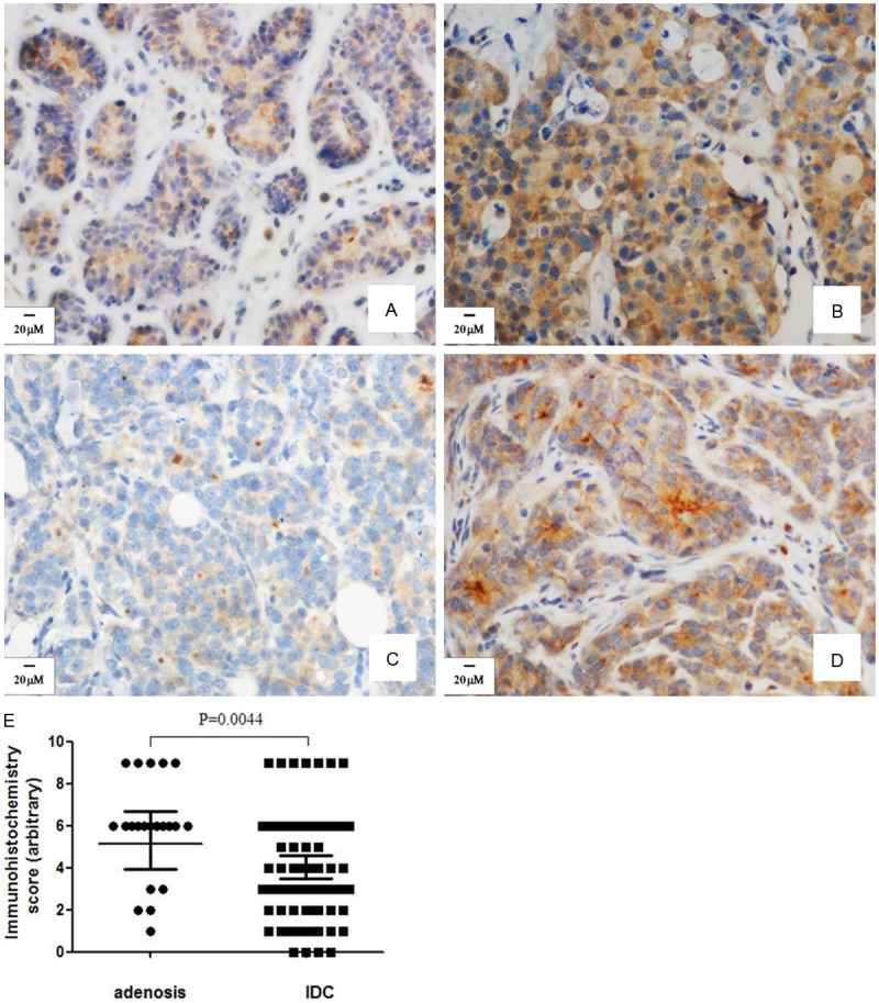
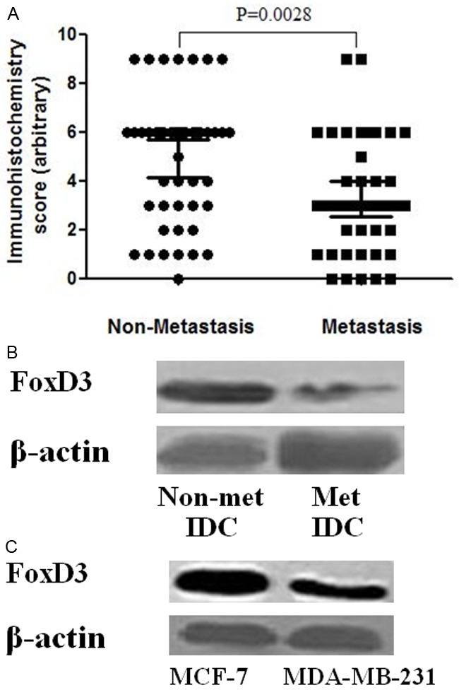
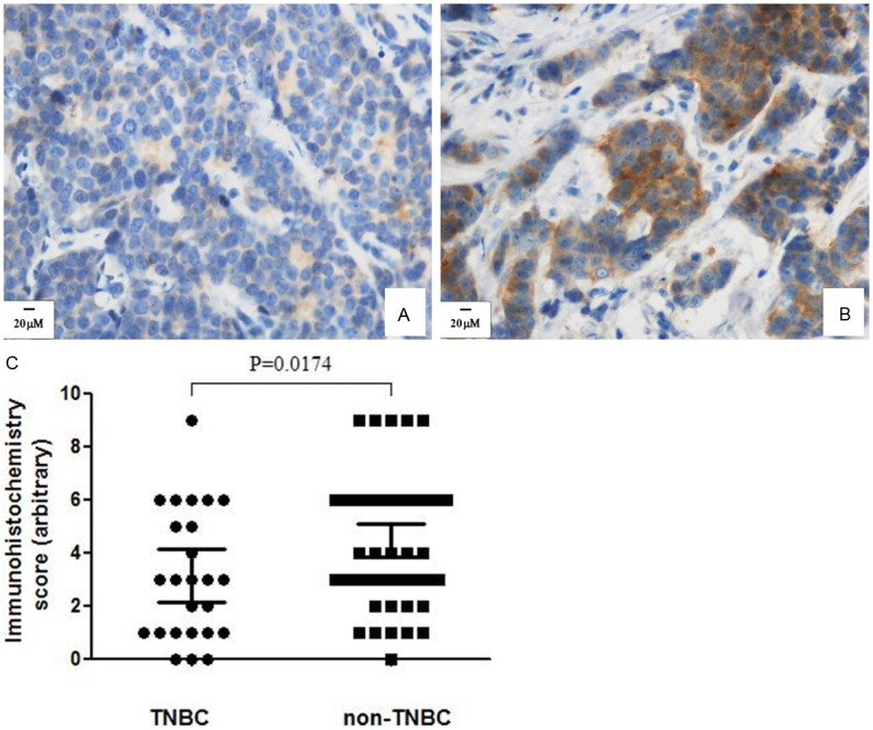
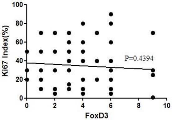
FoxD3 is a transcription factor of the forkhead gene family. We investigated its expression in invasive ductal carcinomas (IDC) of the breast and its association with metastasis. The expression of FoxD3, human epidermal growth factor receptor-2 (HER-2), estrogen receptor (ER), progesterone receptor (PR) and Ki67 was examined by immunohistochemistry in samples from 121 patients with IDC. Non-tumorous breast adenosis tissues served as controls. HER2 expression was confirmed by fluorescence in situ hybridization (FISH). The expression levels of FoxD3 in IDC tissues and the breast cancer cell lines MCF-7 and MDA-MB-231 were additionally measured by western blotting. A greater percentage of total IDC patients and patients with lymph node metastases showed reduced FoxD3 expression compared to adenosis controls (p<0.05). Overall, FoxD3 was associated with metastatic status of IDC but not with age, pathological or clinical staging, or status of HER-2, ER, or PR. In particular, FoxD3 protein expression was down-regulated in the tumor epithelia of IDC samples from patients with metastases. Furthermore, FoxD3 protein expression was decreased in the metastatic MDA-MB-231 breast cancer cell line relative to the non-metastatic cell line, MCF-7. A greater number of patients with invasive, triple-negative breast cancer were also negative for FoxD3 expression than in other, non-triple-negative tumor types. These results suggest an inverse relationship between FoxD3 expression and tumor metastasis and warrants further investigation.
Keywords: Breast cancer; FoxD3; HER-2; invasive ductal carcinomas.
Figures
Similar articles
-
Relation between Ki-67, ER, PR, Her2/neu, p21, EGFR, and TOP II-α expression in invasive ductal breast cancer patients and correlations with prognosis.Asian Pac J Cancer Prev. 2015;16(2):823-9. doi: 10.7314/apjcp.2015.16.2.823. Asian Pac J Cancer Prev. 2015. PMID: 25684532
-
Alterations of estrogen receptors, progesterone receptors and c-erbB2 oncogene protein expression in ductal carcinomas of the breast.Cell Biol Int. 2008 Jun;32(6):698-707. doi: 10.1016/j.cellbi.2008.01.007. Epub 2008 Jan 25. Cell Biol Int. 2008. PMID: 18296077
-
GATA3 expression in breast carcinoma: utility in triple-negative, sarcomatoid, and metastatic carcinomas.Hum Pathol. 2013 Jul;44(7):1341-9. doi: 10.1016/j.humpath.2012.11.003. Epub 2013 Jan 31. Hum Pathol. 2013. PMID: 23375642 Free PMC article.
-
Impact of molecular subtypes classification concordance between preoperative core needle biopsy and surgical specimen on early breast cancer management: Single-institution experience and review of published literature.Eur J Surg Oncol. 2017 Apr;43(4):642-648. doi: 10.1016/j.ejso.2016.10.025. Epub 2016 Nov 17. Eur J Surg Oncol. 2017. PMID: 27889196 Review.
-
Potential prognostic tumor biomarkers in triple-negative breast carcinoma.Beijing Da Xue Xue Bao Yi Xue Ban. 2012 Oct 18;44(5):666-72. Beijing Da Xue Xue Bao Yi Xue Ban. 2012. PMID: 23073572 Review.
Cited by
-
Epithelial-to-mesenchymal transition in thyroid cancer: a comprehensive review.Endocrine. 2019 Dec;66(3):435-455. doi: 10.1007/s12020-019-02030-8. Epub 2019 Aug 4. Endocrine. 2019. PMID: 31378850 Review.
-
FOXD3 regulates anaplastic thyroid cancer progression.Oncotarget. 2017 May 16;8(20):33644-33651. doi: 10.18632/oncotarget.16853. Oncotarget. 2017. PMID: 28430585 Free PMC article.
-
FOXD3 may be a new cellular target biomarker as a hypermethylation gene in human ovarian cancer.Cancer Cell Int. 2019 Feb 28;19:44. doi: 10.1186/s12935-019-0755-8. eCollection 2019. Cancer Cell Int. 2019. PMID: 30858761 Free PMC article.
-
Enhanced JunD/RSK3 signalling due to loss of BRD4/FOXD3/miR-548d-3p axis determines BET inhibition resistance.Nat Commun. 2020 Jan 14;11(1):258. doi: 10.1038/s41467-019-14083-4. Nat Commun. 2020. PMID: 31937753 Free PMC article.
-
Chromatin accessibility landscape and active transcription factors in primary human invasive lobular and ductal breast carcinomas.Breast Cancer Res. 2022 Jul 29;24(1):54. doi: 10.1186/s13058-022-01550-y. Breast Cancer Res. 2022. PMID: 35906698 Free PMC article.
References
-
- Jemal A, Bray F, Center MM, Ferlay J, Ward E, Forman D. Global cancer statistics. CA Cancer J Clin. 2011;61:69–90. - PubMed
-
- Ezeh UI, Turek PJ, Reijo RA, Clark AT. Human embryonic stem cell genes OCT4, NANOG, STELLAR, and GDF3 are expressed in both seminoma and breast carcinoma. Cancer. 2005;104:2255–2265. - PubMed
-
- Sutton J, Costa R, Klug M, Field L, Xu D, Largaespada DA, Fletcher CF, Jenkins NA, Copeland NG, Klemsz M, Hromas R. Genesis, a winged helix transcriptional repressor with expression restricted to embryonic stem cells. J Biol Chem. 1996;271:23126–23133. - PubMed
Publication types
MeSH terms
Substances
LinkOut - more resources
Full Text Sources
Medical
Research Materials
Miscellaneous