

Plexiform angiomyxoid myofibroblastic tumor (PAMT) of the stomach. A case report focusing on its characteristic growth pattern
- PMID: 24551290
- PMCID: PMC3925914
Plexiform angiomyxoid myofibroblastic tumor (PAMT) of the stomach. A case report focusing on its characteristic growth pattern
Abstract
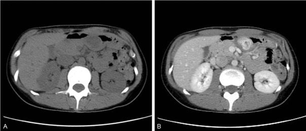
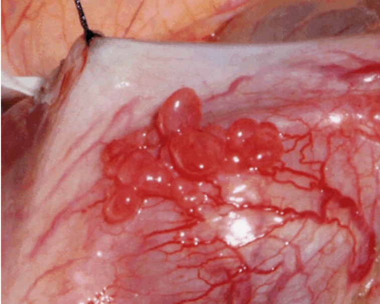
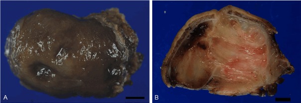
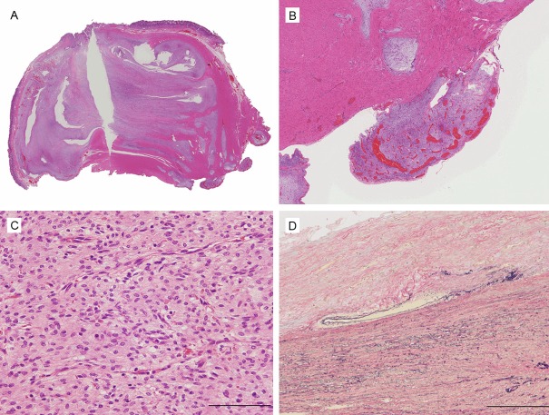
Plexiform angiomyxoid myofibroblastic tumor (PAMT) is a rare mesenchymal tumor of the stomach. We report herein a case with CT findings, which illustrate the characteristic growth pattern of PAMT. A 27-year-old female patient visited our hospital because of epigastric pain and anemia. The CT scan showed a heterogeneous tumor in the gastric antrum, which was drastically enhanced with contrast medium, and consisted of a number of highly stained small nodules around the tumor rim. The resected tumor, 4.6 cm in size, was c-kit negative and SMA-positive by immunohistochemistry, and composed of bland spindle cells which were separated by abundant myxomatous stroma. The tumor showed plexiform growth in the entire stomach wall, with multiple nodules protruding outward within the serosa. The CT findings in this case reflect the characteristic PAMT growth pattern, and are distinct enough to differentiate it from gastrointestinal stromal tumor (GIST).
Keywords: CT; GIST; PAMT; diagnosis.
Figures
Similar articles
-
Plexiform angiomyxoid myofibroblastic tumor of the stomach: A case report.Diagn Cytopathol. 2017 Jan;45(1):55-58. doi: 10.1002/dc.23572. Epub 2016 Aug 26. Diagn Cytopathol. 2017. PMID: 27561459
-
Plexiform angiomyxoid myofibroblastic tumor of the stomach: a case report.J Korean Med Sci. 2011 Nov;26(11):1508-11. doi: 10.3346/jkms.2011.26.11.1508. Epub 2011 Oct 27. J Korean Med Sci. 2011. PMID: 22065909 Free PMC article.
-
Presence of smooth muscle cell differentiation in plexiform angiomyxoid myofibroblastic tumor of the stomach: a case report.Int J Clin Exp Pathol. 2014 Jan 15;7(2):823-7. eCollection 2014. Int J Clin Exp Pathol. 2014. PMID: 24551311 Free PMC article.
-
[Plexiform angiomyxoid myofibroblastic tumor of stomach].Zhonghua Bing Li Xue Za Zhi. 2012 Nov;41(11):756-60. doi: 10.3760/cma.j.issn.0529-5807.2012.11.010. Zhonghua Bing Li Xue Za Zhi. 2012. PMID: 23302337 Review. Chinese.
-
Plexiform angiomyxoid myofibroblastic tumor treated by endoscopic submucosal dissection: A case report and review of the literature.World J Gastroenterol. 2021 Aug 21;27(31):5288-5296. doi: 10.3748/wjg.v27.i31.5288. World J Gastroenterol. 2021. PMID: 34497451 Free PMC article. Review.
Cited by
-
Imaging findings of gastric plexiform fibromyxoma with a cystic change: A case report and review of literature.Medicine (Baltimore). 2017 Dec;96(52):e8967. doi: 10.1097/MD.0000000000008967. Medicine (Baltimore). 2017. PMID: 29384895 Free PMC article. Review.
-
Unusual focal keratin expression in plexiform angiomyxoid myofibroblastic tumor: A case report and review of the literature.Medicine (Baltimore). 2016 Jul;95(28):e4207. doi: 10.1097/MD.0000000000004207. Medicine (Baltimore). 2016. PMID: 27428222 Free PMC article. Review.
-
Plexiform fibromyxoma of the small bowel: A case report.World J Clin Cases. 2018 Dec 6;6(15):1067-1072. doi: 10.12998/wjcc.v6.i15.1067. World J Clin Cases. 2018. PMID: 30568965 Free PMC article.
-
An Update on Clinicopathological and Molecular Features of Plexiform Fibromyxoma.Can J Gastroenterol Hepatol. 2019 Jul 7;2019:3960920. doi: 10.1155/2019/3960920. eCollection 2019. Can J Gastroenterol Hepatol. 2019. PMID: 31360694 Free PMC article. Review.
-
Plexiform fibromyxoma: a clinicopathological and immunohistochemical analysis of two cases with a literature review.J Int Med Res. 2021 Aug;49(8):3000605211027878. doi: 10.1177/03000605211027878. J Int Med Res. 2021. PMID: 34369189 Free PMC article. Review.
References
-
- Takahashi Y, Shimizu S, Ishida T, Aita K, Toida S, Fukusato T, Mori S. Plexiform angiomyxoid myofibroblastic tumor of the stomach. Am J Surg Pathol. 2007 May;31:724–728. - PubMed
-
- Galant C, Rousseau E, Ho Minh Duc DK, Pauwels P. Plexiform angiomyxoid myofibroblastic tumor of the stomach. Am J Surg Pathol. 2008 Dec;32:1910. - PubMed
-
- Yoshida A, Klimstra DS, Antonescu CR. Plexiform angiomysoid tumor of the stomach. Am J Surg Pathol. 2008 Dec;32:1910–1912. - PubMed
-
- Miettinen M, Makhlouf HR, Sobin LH, Lasota J. Plexiform Fibromyxoma. Am J Surg Pathol. 2009 Nov;33:1624–1632. - PubMed
-
- Rau TT, Hartmann A, Dietmaier W, Schmitz J, Hohenberger W, Hofstaedter F, Katenkamp K. Plexiform angiomyxoid myofibroblastic tumour: differential diagnosis of gastrointestinal stromal tumour in the stomach. J Clin Pathol. 2008;61:1136–1137. - PubMed
Publication types
MeSH terms
Substances
LinkOut - more resources
Full Text Sources
Medical