

In vitro ovarian cancer model based on three-dimensional agarose hydrogel
- PMID: 24551446
- PMCID: PMC3924902
- DOI: 10.1177/2041731413520438
In vitro ovarian cancer model based on three-dimensional agarose hydrogel
Abstract
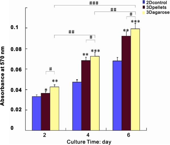
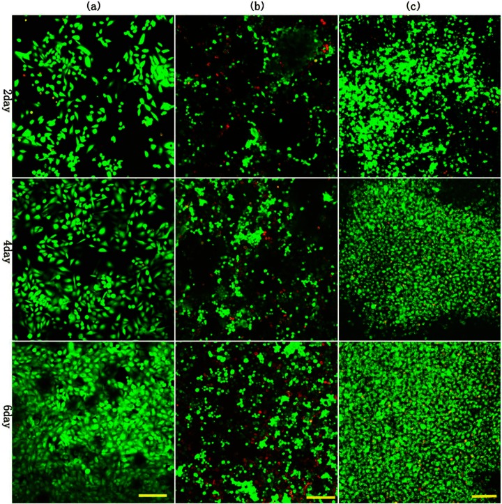
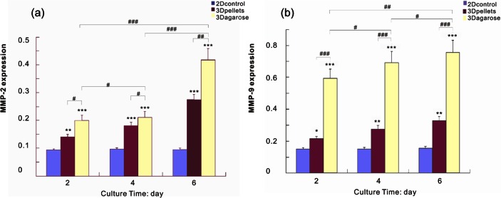
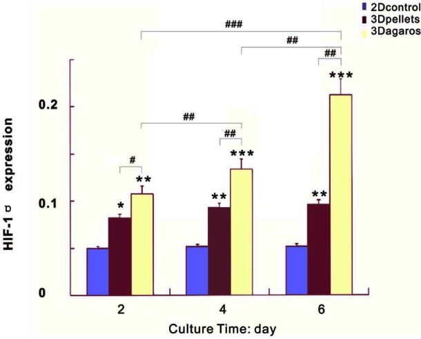
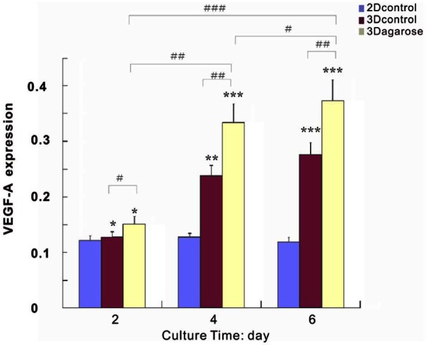
To establish a typical tumor model of ovarian cancer which may be more representative and reliable than traditional monolayer culture and pellet, agarose was used as cell vehicle to engineering tumor. Selection of agarose is based on its successful application in tissue engineering with both amenable mechanical and biological properties. In this study, ovarian cancer cell line SKOV3 was encapsulated in agarose hydrogel with cell aggregates and two-dimensional culture as controls. In vitro cell proliferation was assessed by MTT and cell viability was examined at time points of 2, 4, and 6 days. The expression of tumor malignancy markers including matrix metalloproteinase 2, matrix metalloproteinase 9, hypoxia-inducible factor-1α, and vascular endothelial growth factor-A was assessed by real-time polymerase chain reaction. The results showed that cells proliferated more rapidly in three-dimensional agarose culture than controls. Furthermore, upregulation of matrix metalloproteinase 9 and matrix metalloproteinase 2 activity and increased expression of vascular endothelial growth factor-A and hypoxia-inducible factor-1α were shown in agarose-engineered tumors. All the evidences demonstrated that agarose may provide a more favorable environment for cancer cell growth, mimicking the in vivo environment for tumor generation. The novel in vitro tumor model may be useful for the further investigation of anticancer therapeutics.
Keywords: Agarose; ovarian cancer; tumor engineering; tumor microenvironment.
Conflict of interest statement
Figures

Similar articles
-
In vivo bioengineered ovarian tumors based on collagen, matrigel, alginate and agarose hydrogels: a comparative study.Biomed Mater. 2015 Jan 29;10(1):015016. doi: 10.1088/1748-6041/10/1/015016. Biomed Mater. 2015. PMID: 25634132
-
Engineering three-dimensional constructs of the periodontal ligament in hyaluronan-gelatin hydrogel films and a mechanically active environment.J Periodontal Res. 2013 Dec;48(6):790-801. doi: 10.1111/jre.12072. Epub 2013 Apr 15. J Periodontal Res. 2013. PMID: 23581542
-
In vitro maturation on ovarian granulosa cells encapsulated in agarose matrix improves developmental competence of porcine oocytes.Theriogenology. 2021 Apr 1;164:42-50. doi: 10.1016/j.theriogenology.2021.01.008. Epub 2021 Jan 21. Theriogenology. 2021. PMID: 33540369
-
Cardiac-Derived Extracellular Matrix Enhances Cardiogenic Properties of Human Cardiac Progenitor Cells.Cell Transplant. 2016;25(9):1653-1663. doi: 10.3727/096368915X689794. Epub 2015 Nov 16. Cell Transplant. 2016. PMID: 26572770
-
An HCG-rich microenvironment contributes to ovarian cancer cell differentiation into endothelioid cells in a three-dimensional culture system.Oncol Rep. 2015 Nov;34(5):2395-402. doi: 10.3892/or.2015.4215. Epub 2015 Aug 20. Oncol Rep. 2015. PMID: 26479853
Cited by
-
Recent advances in 3D bioprinted tumor models for personalized medicine.Transl Oncol. 2023 Nov;37:101750. doi: 10.1016/j.tranon.2023.101750. Epub 2023 Aug 10. Transl Oncol. 2023. PMID: 37572498 Free PMC article.
-
Biocompatible micro tweezers for 3D hydrogel organoid array mechanical characterization.PLoS One. 2022 Jan 24;17(1):e0262950. doi: 10.1371/journal.pone.0262950. eCollection 2022. PLoS One. 2022. PMID: 35073389 Free PMC article.
-
Cancer Cell Direct Bioprinting: A Focused Review.Micromachines (Basel). 2021 Jun 28;12(7):764. doi: 10.3390/mi12070764. Micromachines (Basel). 2021. PMID: 34203530 Free PMC article. Review.
-
Scaffold Using Chitosan, Agarose, Cellulose, Dextran and Protein for Tissue Engineering-A Review.Polymers (Basel). 2023 Mar 19;15(6):1525. doi: 10.3390/polym15061525. Polymers (Basel). 2023. PMID: 36987305 Free PMC article. Review.
-
Development of a hydrogel-based three-dimensional (3D) glioblastoma cell lines culture as a model system for CD73 inhibitor response study.Biomed Eng Online. 2024 Dec 21;23(1):127. doi: 10.1186/s12938-024-01320-1. Biomed Eng Online. 2024. PMID: 39709472 Free PMC article.
References
LinkOut - more resources
Full Text Sources
Other Literature Sources
