. 2014 Feb 5:4:11.
doi: 10.3389/fcimb.2014.00011. eCollection 2014.

A murine oral model for Mycobacterium avium subsp. paratuberculosis infection and immunomodulation with Lactobacillus casei ATCC 334

Affiliations

A murine oral model for Mycobacterium avium subsp. paratuberculosis infection and immunomodulation with Lactobacillus casei ATCC 334

Meagan A Cooney et al. Front Cell Infect Microbiol. .

Abstract

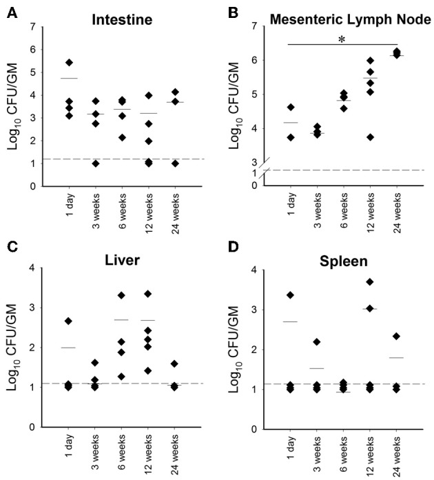
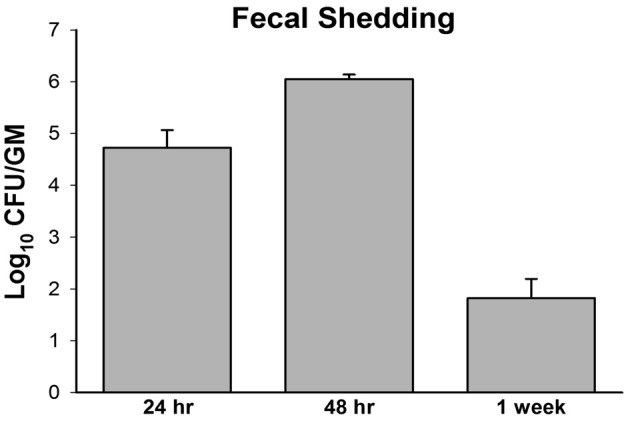
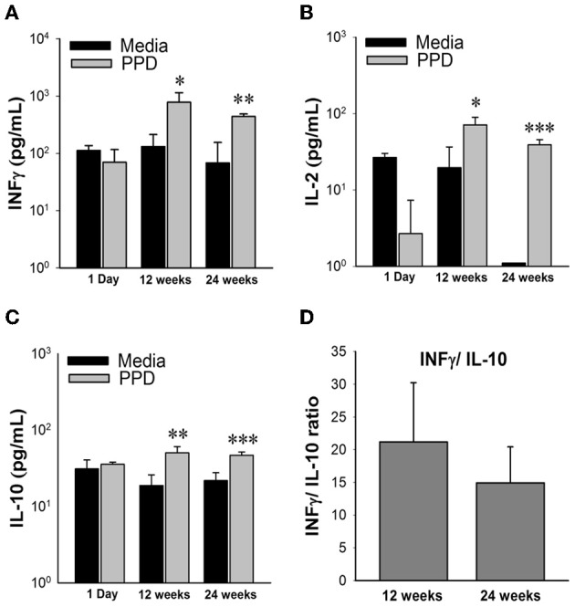
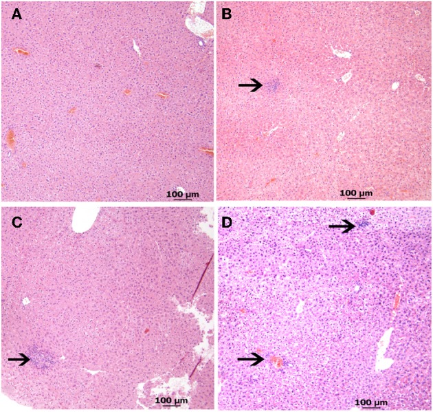
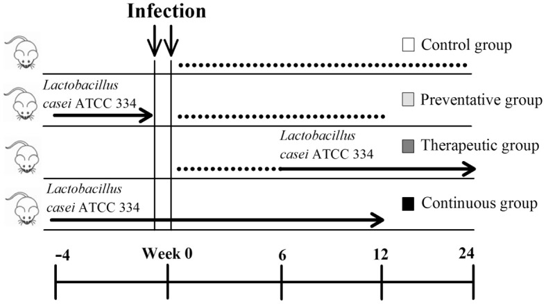
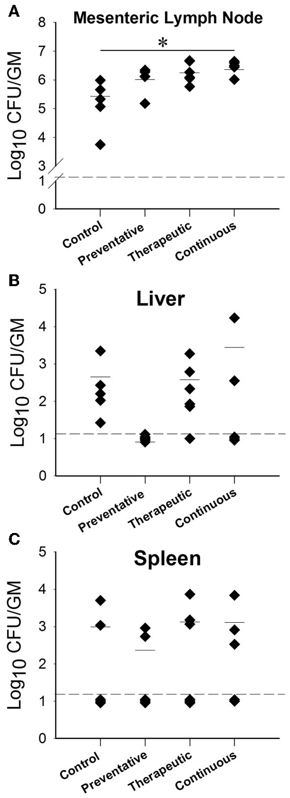
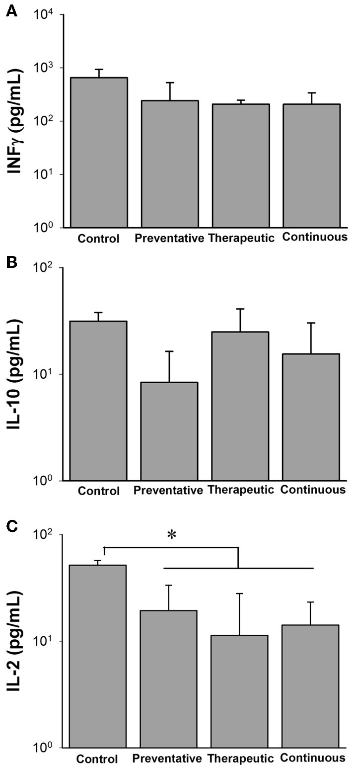
Mycobacterium avium subsp. paratuberculosis (M. paratuberculosis) the causative agent of Johne's disease, is one of the most serious infectious diseases in dairy cattle worldwide. Due to the chronic nature of this disease and no feasible control strategy, it is essential to have an efficient animal model which is representative of the natural route of infection as well as a viable treatment option. In this report, we evaluated the effect of different doses of M. paratuberculosis in their ability to colonize murine tissues following oral delivery and the ability of Lactobacillus casei ATCC 334, a nascent probiotic, to combat paratuberculosis. Oral inoculation of mice was able to establish paratuberculosis in a dose-dependent manner. Two consecutive doses of approximately 10(9) CFU per mouse resulted in a disseminated infection, whereas lower doses were not efficient to establish infection. All inoculated mice were colonized with M. paratuberculosis, maintained infection for up to 24 weeks post infection and generated immune responses that reflect M. paratuberculosis infection in cattle. Notably, oral administration of L. casei ATCC 334 did not reduce the level of M. paratuberculosis colonization in treated animals. Interestingly, cytokine responses and histology indicated a trend for the immunomodulation and reduction of pathology in animals receiving L. casei ATCC 334 treatment. Overall, a reproducible oral model of paratuberculosis in mice was established that could be used for future vaccine experiments. Although the L. casei ATCC 334 was not a promising candidate for controlling paratuberculosis, we established a protocol to screen other probiotic candidates.

Keywords: animal model; oral model; pratuberculosis; probiotics; vaccine development.


Figures

Figure 1
Figure 1
Organ colonization from high dose orally infected mice. Graphs (A–D) (A, Intestine; B, Mesenteric lymph node; C, Liver; D, Spleen) depict CFU/g obtained from plating serial dilutions of homogenized organs (intestines, mesenteric lymph node, liver and spleen) from mice orally infected with two consecutive doses of 109 CFU of M. avium subsp. paratuberculosis K10 over a 24 week period. Each dot represents one mouse, the hash mark denotes the mean, and the dotted line represents the minimum detection limit. (*p < 0.05).
Figure 2
Figure 2
M. avium subsp. paratuberculosis shedding in mouse fecal samples. Pooled fecal samples were collected from mice orally infected with two consecutive doses of 109 CFU of M. avium subsp. paratuberculosis K10 Samples were decontaminated by HPC treatment and serial dilution plating on 7H10 agar was performed for quantification. All samples were run in duplicate.
Figure 3
Figure 3
Cytokine profiles of high dose M. avium subsp. paratuberculosis orally infected mice over 24 weeks post infection. Graphs (A–C) depict cytokine levels of mice orally infected with two consecutive doses of 109 CFU of M. avium subsp. paratuberculosis K10 over a 24 week period. At time of sacrifice, mouse spleens were collected and splenocytes were isolated and stimulated with Johnin Purified Protein Derivative (PPD) or media only for 48 h. Supernatant was collected and used for cytokine quantification by luminex bead array. Black bars represent the mean levels of media only background levels with their standard deviation and gray bars represent the mean levels from stimulation with PPD with their standard deviation. Significant increases in PPD stimulated splenocytes were determined by comparison to media only levels. (*p < 0.05, **p < 0.01, ***p < 0.001). To determine shifts in immune response from 12 to 24 weeks, levels of IFNγ were divided by IL-10 from individual mice (D). The mean ratio and standard deviation for each time point is shown.
Figure 4
Figure 4
Liver pathology from high dose oral M. avium subsp. paratuberculosis K10 challenge at various time points. Images (A–D) reveal liver pathology from mice orally infected with two consecutive doses of 109 CFU of M. avium subsp. paratuberculosis K10 over a 24 week period. H&E stained sections with 40 × magnification (scale bar = 100 μm) are shown. Images of 6 and 12 weeks post infection show lymphocyte inflammation and the image at 24 weeks post infection shows granulomatous inflammation. (A) Non-infected (B) 6 weeks (C) 12 weeks (D) 24 weeks.
Figure 5
Figure 5
Experimental design of Lactobacillus casei ATCC 334 feedings. Three Lactobacillus casei ATCC 334 trials were conducted (preventative, therapeutic and continuous) along with a control. All mice were inoculated with two doses of 109 CFU of M. avium subsp. paratuberculosis K10 on two consecutive days. The blue arrows represent the period of ATCC 334 feeding, where mice received daily doses of ~109 CFU of ATCC 334. The black lines denote the parts of the trial period where no ATCC 334 was given. Mice were sacrificed and samples for colonization, histopathology and immunology were collected at 12 and/or 24 weeks post infection.
Figure 6
Figure 6
M. avium subsp. paratuberculosis organ colonization from different Lactobacillus casei ATCC 334 treatments at 12 weeks post oral M. avium subsp. paratuberculosis K10 infection. Graphs (A–C) (A-Mesenteric lymph node, B-Liver, C-Spleen) depict the CFU/g of M. avium subsp. paratuberculosis obtained from the homogenized organs (mesenteric lymph node, liver and spleen) of mice treated with different ATCC 334 treatments and orally challenged with two doses of 109 CFU M. avium subsp. paratuberculosis K10. Each dot represents one mouse, the hash mark denotes the mean, and the dotted line represents the minimum detection limit. (*p < 0.05).
Figure 7
Figure 7
Cytokine profiles of different Lactobacillus casei ATCC 334 treatments at 12 weeks post oral M. avium subsp. paratuberculosis K10 infection. Graphs A–C (A. INFγ, B. IL-10, C. IL-12) depict cytokine levels of mice orally infected with two consecutive doses of 109 CFU of M. avium subsp. paratuberculosis K10 and treated with different ATCC 334 treatments. At 12 weeks post infection, mouse spleens were collected and splenocytes were isolated and stimulated with Johnin Purified Protein Derivative (PPD) or media only for 48 h. Supernatant was collected and used for cytokine quantification by luminex bead array. Graph bars reflect the mean of each treatment group and their standard deviation. Levels were calculated by subtracting individual PPD stimulated levels from their background levels. Significant differences among groups are reflected by *p < 0.05.

References

    1. Chacon O., Bermudez L. E., Barletta R. G. (2004). Johne's disease, inflammatory bowel disease, and Mycobacterium paratuberculosis. Annu. Rev. Microbiol. 58, 329–363 10.1146/annurev.micro.58.030603.123726 - DOI - PubMed
    1. Chamberlin W., Graham D. Y., Hulten K., El-Zimaity H. M. T., Schwartz M. R., Shafran I., et al. (2001). Review article: Mycobacterium avium subsp. paratuberculosis as one cause of Crohn's disease. Aliment. Pharmacol. Ther. 15, 337–346 10.1046/j.1365-2036.2001.00933.x - DOI - PubMed
    1. Collins M. T. (2011). Diagnosis of paratuberculosis. Vet. Clin. North Am. Food Anim. Pract. 27 581–591, vi. 10.1016/j.cvfa.2011.07.013 - DOI - PubMed
    1. Corpa J. M., Perez V., Sanchez M. A., Marin J. F. (2000). Control of paratuberculosis (Johne's disease) in goats by vaccination of adult animals. Vet. Rec. 146, 195–196 10.1136/vr.146.7.195 - DOI - PubMed
    1. Corr S. C., Hill C., Gahan C. G. M. (2009). Understanding the mechanisms by which probiotics inhibit gastrointestinal pathogens, in Advances in Food and Nutrition Research, Vol. 56, ed Taylor S. L. (Academic Press; ), 1–15 10.1016/S1043-4526(08)00601-3 - DOI - PubMed

Publication types

LinkOut - more resources