. 2014 Jun;16(6):797-815.
doi: 10.1111/cmi.12279. Epub 2014 Mar 21.

Mycobacterium leprae intracellular survival relies on cholesterol accumulation in infected macrophages: a potential target for new drugs for leprosy treatment

Affiliations
Free PMC article

Mycobacterium leprae intracellular survival relies on cholesterol accumulation in infected macrophages: a potential target for new drugs for leprosy treatment

Katherine A Mattos et al. Cell Microbiol. 2014 Jun.
Free PMC article

Abstract

We recently showed that Mycobacterium leprae (ML) is able to induce lipid droplet formation in infected macrophages. We herein confirm that cholesterol (Cho) is one of the host lipid molecules that accumulate in ML-infected macrophages and investigate the effects of ML on cellular Cho metabolism responsible for its accumulation. The expression levels of LDL receptors (LDL-R, CD36, SRA-1, SR-B1, and LRP-1) and enzymes involved in Cho biosynthesis were investigated by qRT-PCR and/or Western blot and shown to be higher in lepromatous leprosy (LL) tissues when compared to borderline tuberculoid (BT) lesions. Moreover, higher levels of the active form of the sterol regulatory element-binding protein (SREBP) transcriptional factors, key regulators of the biosynthesis and uptake of cellular Cho, were found in LL skin biopsies. Functional in vitro assays confirmed the higher capacity of ML-infected macrophages to synthesize Cho and sequester exogenous LDL-Cho. Notably, Cho colocalized to ML-containing phagosomes, and Cho metabolism impairment, through either de novo synthesis inhibition by statins or depletion of exogenous Cho, decreased intracellular bacterial survival. These findings highlight the importance of metabolic integration between the host and bacteria to leprosy pathophysiology, opening new avenues for novel therapeutic strategies to leprosy.


Figures

Fig 1
Fig 1
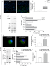
Lipid accumulation in vivo and in vitro in ML-infected tissues and cells.A. Sections of skin biopsies from BT and LL patients (n = 4) were immunostained for ADRP and observed by immunofluorescence confocal microscopy. Bar: 20 μm (yellow).B. Total RNA was extracted from BT and LL biopsies, and expression of ADRP was normalized to GADPH and quantified by qRT-PCR. Values are the average of four independent experiments.C. Representative HPTLC of neutral lipids extracted from skin biopsies of BT and LL patients detected by charring.D. The content of each class of lipids was estimated by densitometry and plotted as a percentage.E. Lipid accumulation in uninfected and ML-infected macrophages was observed by immunostaining for ADRP. Bar: 10 μm (white).F. LD formation was measured by Nile Red staining.G and H. (G) Representative HPTLC of neutral lipids extracted from macrophages treated or not with ML and (H) the percentage of each class of lipids estimated by densitometry.I. Quantitative Cho levels estimated by filipin staining and flow cytometry.In (A) and (E) Nuclei (blue) were labelled with DAPI. No fluorescence was observed for the Alexa546-labelled mouse isotype control IgG. In HPTLC, the position of pure standard lipids is indicated on the margin of the panel. *Statistically significant differences (P ≤ 0.05) comparing LL groups, BT groups, and ML-infected cells versus uninfected cells.
Fig 2
Fig 2
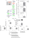
Modulation of host steroid metabolism during ML infection.A. Steroid biosynthetic pathway in humans. Metabolites present at different levels in BT and LL samples are coloured red (higher in BT samples) and green (higher in LL samples), and their m/z values are indicated within parentheses.B. Signal intensity values of metabolites indicated in (A). White columns indicate average signal intensities from BT samples whereas grey columns indicate average intensity values from LL samples. Bars indicate the standard errors of means.C and D. HMGCR expression in leprosy skin biopsies. (C) Total RNA was extracted from BT and LL biopsies and the expression of HMGCR was quantified by qRT-PCR, being normalized to GADPH levels. Values are the average of four independent experiments. (D) Cell lysates (30 μg of protein) were subjected to SDS-PAGE and analysed by Western blot with anti-HMGCR and anti-β-tubulin antibodies. The figure shows a representative Western blot. The graph shows the normalized values (tubulin) from three independent experiments.E. Total RNA was isolated from BT and LL skin biopsies and subjected to qRT-PCR analysis to measure the enzymes from the de novo Cho pathway as shown in (A).F. Acetate incorporation was assayed in uninfected and ML-infected macrophages after 48 h. Incorporation of [3H] acetate into the cholesterol (Cho) and cholesteryl ester (ChoE) was measured in counts per minute of [3H] acetate incorporated into 106 macrophages and expressed as percentages relative to the control (uninfected cells) ± SEM from three separate experiments.Statistically significant differences were at P ≤ 0.05 when the BT group was compared to the LL group and uninfected cultures to ML-infected cells. *Statistically significant differences (P ≤ 0.05) comparing LL with BT. Statistically significant differences (P ≤ 0.05) comparing ML-stimulated versus control cells.
Fig 3
Fig 3
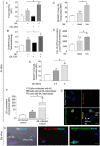
Cellular incorporation of LDL-cholesterol in ML-infected macrophages. Forty-eight hours after infection in medium containing 2% FCS, infected macrophages were pulse-labelled at 33°C with LDL-Cho for 2 h in medium serum free.A. The effect of native LDL-Cho and ML infection on intracellular Cho levels measured by filipin staining and flow cytometry.B. In parallel, the quantitative analysis of total Cho was determined by Amplex Red Cholesterol kit under the same conditions.C. Cells were treated with dead or live bacteria in the same condition described above and the LDL-[BODIPY-Cho] incorporation was determined by flow cytometry.D. To validate the fluorescence assay the LDL-[3H]-Cho incorporation was assayed under same condition of fluorescence assay.C–E. The internalization of LDL-[BODIPY-Cho] as a process dependent on bacterial viability (C and D) and multiplicity of infection (moi) (E) as determined by flow cytometry.F. Bacterial association and LDL-[BODIPY-Cho] were measured simultaneously by flow cytometry. A separate analysis was performed in cells with no bacteria (cells without internalized ML) and cells bearing bacteria (cells with internalized ML). MFI values of LDL-[BODIPY-Cho] are expressed in bar graphs.Results from five representative experiments are shown. *Statistically significant differences (P ≤ 0.05) when comparing ML-infected cells with control cells. +Statistically significant differences (P ≤ 0.05) when comparing different ML-treated cell groups; n.s: non-significant.G and H. Fluorescence images of macrophage cultures showing LDL-[BODIPY-Cho] in cells associated or not with ML. (G) Macrophages isolated from PBMC and (H) Virchow cells isolated from LL skin biopsies. Original bar, 10 μm.
Fig 4
Fig 4
LDL receptors are upregulated by ML infection of human macrophages.A. Total RNA was isolated from LL and BT skin biopsies and subjected to qRT-PCR analysis to measure LDLR, LRP-1, SR-A1, SR-B1, CD-36 and GADPH mRNA expression. Values are the average of four independent experiments.B. Leprosy lesions were labelled with the monoclonal antibody SR-A1. Shown are representative sections from LL and BT lesions. In BT skin lesions the distribution of few SR-A1+ cells was concentrated in inflammatory infiltrates and nearly to epidermis (arrows). LL skin lesions presented a higher positive SR-A1 cells within the dermis, mainly in inflammatory infiltrates (arrows). Photomicrographs are representative of four experiments performed. Scale bar: 50 μm.C and D. Cell lysates (20 μg of protein) were subjected to SDS-PAGE and analysed by Western blot with anti-LDL-R (C), anti-CD36 (D) and as loading control, anti-GADPH antibodies (C–D). The figure shows a representative Western blot. The graph shows the normalized values (GADPH) from three independent experiments.E and F. (E) CD36 expression in human macrophages and (F) WT and TLR2−/− murine macrophages was performed using flow cytometry.*P ≤ 0.05, ML-stimulated versus control cells and LL versus BT groups. +P ≤ 0.05 between the different ML-treated cell groups.
Fig 5
Fig 5
ML in vivo infection induces SREBP expression and activation.A. The expression of SREBPs was compared in skin biopsies of LL versus BT of leprosy patients by qRT-PCR.B and C. Total RNA was extracted from BT and LL biopsies and the expression of SREBP was quantified by qRT-PCR, being normalized to loading controls α-tubulin (B) and GADPH (C) levels. The levels of the activated/mature forms (mSREBP) of both SREBP-1 (B) and SREBP-2 (C) were analysed by Western blot.The figure shows a representative Western blot. The graph shows the normalized values (loading control) from three independent experiments. *P ≤ 0.05.
Fig 6
Fig 6
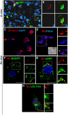
Intracellular cholesterol sources colocalize with ML in foamy macrophages of LL patients.A. Sections of skin biopsies of LL patients (n = 4) were analysed showing colocalization between LD organelles (green) and ML (red).B. High magnification of the boxed areas visualized in (A) showing bacteria completely enveloped by LDs (yellow arrow).C–H. Virchow's cells isolated from LL biopsies were stained with different fluorophores and anti-ADRP to visualize Cho sources within these ML-infected foamy macrophages. (C) Oil red O reveals the foamy phenotype of the isolated Virchow's cell. (D) Fluorescence and (E) Differential interference contrast (DIC) of a Virchow's cell showing ML residing in Cho-rich regions (yellow arrow). (F) LD–ML association within macrophages isolated from LL biopsies was visualized using BODIPY label. (G) Immunofluorescence confocal images for ADRP (green) and anti-LAM (red) show the LD–ML association (yellow arrow) (H) LDL-[BODIPY-Cho] internalization in Virchow's cells and intimate association with ML (yellow arrow). Nuclei were stained with TO-PRO-3. No fluorescence was observed with the isotype control IgG (Supplementary Fig. S2). Bars: white = 10 mm; yellow = 5 mm. Filipin was used as a Cho label (blue). BODIPY and ADRP were used as LD marker (green); LDL-Cho: LDL fluorescent as LDL-[BODIPY-Cho] (green), LAM (lipoarabinomannan) was used as an ML marker (red).
Fig 7
Fig 7
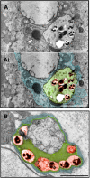
Transmission electron microscopy of a LL skin biopsy showing Mycobacterium leprae-infected macrophages.A and B. Large phagosomes in the macrophage cytoplasm show several bacilli (highlighted in red in Ai and B) enmeshed in a lipid content (highlighted in green in Ai and B). Arrowhead in (A) indicates a typical lipid droplet in association with a phagosome. m, mitochondrium. Bars = 2 μm (A); 1 μm (B).
Fig 8
Fig 8
Intracellular mycobacterial viability is dependent on exogenous and endogenous host cholesterol.A. Phenotype of monocytes isolated from PBMC and infected with ML, treated and not with statin (72 h), was visualized by Oil red O. Bars: 20 mm.B and C. (B) Cho levels extracted from ML-infected monocytes treated or not with statin was determined by HPTLC and by (C) the quantitative analysis determined by Amplex Red Cholesterol kit under the same conditions.D. Monocytes were infected with ML, treated or not with statin, and depleted of LDL from serum (serum-free AIM V medium) or not (RPMI supplemented with FCS) to determine the role of Cho in mycobacterial survival. Percentage of live and dead bacteria was evaluated using the LIVE/DEAD BacLight Bacterial Viability Kit in combination with flow cytometry. Statistically significant (P ≤ 0.05) differences between control and drug-treated groups are indicated by asterisks and + between different drug-treated groups.
Fig 9
Fig 9
A model for host cholesterol acquisition pathways by intracellular ML in macrophages. This figure depicts the potential sources of Cho for intracellular ML. ML infection stimulates the uptake of exogenous Cho sources (LDL) as well as the de novo synthesis of Cho via upregulation of the LDL receptor expression and the HMGCR enzyme, respectively, leading to LD formation. The modulation of Cho metabolism by ML involves the activation of the SREBP-1/2 transcription factors. Internalized LDL and LDs are recruited to bacterial-containing phagosomes where they accumulate. ML modulation of host cell lipid metabolism depends on the TLR2/TLR6 signalling cascade that elicits LD biogenesis and upregulation of CD36. LDs and exogenous lipid recruitment probably constitute an effective ML intracellular strategy to acquire host lipids as a nutritional source and promote bacterial survival. Moreover, accumulation of LDs in infected macrophages favours the generation of an innate immune response with PGE2 production and a high IL-10/IL-12 ratio (Mattos et al., 2010), which may contribute to a permissive environment for ML proliferation. LD: lipid droplet; ML: M. leprae; Cho, cholesterol; ChoE: cholesteryl ester; PGE2: prostaglandin E2; pSREBP: precursor SREBP; mSREBP: mature precursor.

Similar articles

Cited by

References

    1. Adams LB, Soileau NA, Battista JR. Krahenbuhl JL. Inhibition of metabolism and growth of Mycobacterium leprae by gamma irradiation. Int J Lepr Other Mycobact Dis. 2000;68:1–10. - PubMed
    1. Almeida PE, Silva AR, Maya-Monteiro CM, Töröcsik D, D'Avila H, Dezsö B, et al. Mycobacterium bovis bacillus Calmette-Guérin infection induces TLR2-dependent peroxisome proliferator-activated receptor gamma expression and activation: functions in inflammation, lipid metabolism, and pathogenesis. J Immunol. 2009;183:1337–1345. - PubMed
    1. Almeida PE, Roque NR, Magalhães KG, Mattos KA, Teixeira L, Maya-Monteiro C, et al. Differential TLR2 downstream signaling regulates lipid metabolism and cytokine production triggered by Mycobacterium bovis BCG infection. Biochim Biophys Acta. 2014;1841:97–107. - PubMed
    1. Amaral JJ, Antunes LCM, de Macedo CS, Mattos KA, Han J, Pan J, et al. Metabonomics reveals drastic changes in anti-inflammatory/pro-resolving polyunsaturated fatty acids-derived lipid mediators in leprosy disease. PLoS Negl Trop Dis. 2013;7:e2381. doi: 10.1371/journal.pntd.0002381. - DOI - PMC - PubMed
    1. Antunes LC, Han J, Ferreira RB, Lolić P, Borchers CH. Finlay BB. Effect of antibiotic treatment on the intestinal metabolome. Antimicrob Agents Chemother. 2011;55:1494–1503. - PMC - PubMed

Publication types

MeSH terms