. 2014 Mar 6;370(10):911-20.
doi: 10.1056/NEJMoa1307361. Epub 2014 Feb 19.

Early-onset stroke and vasculopathy associated with mutations in ADA2

Affiliations

Early-onset stroke and vasculopathy associated with mutations in ADA2

Qing Zhou et al. N Engl J Med. .

Abstract

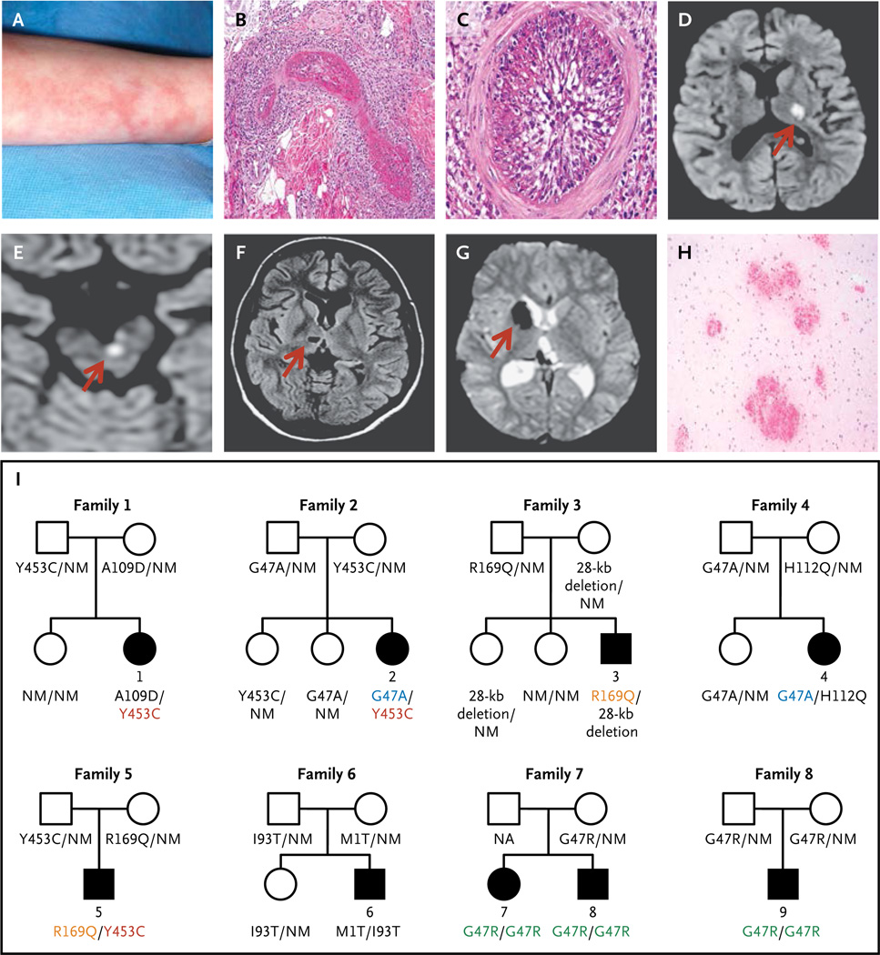
Background: We observed a syndrome of intermittent fevers, early-onset lacunar strokes and other neurovascular manifestations, livedoid rash, hepatosplenomegaly, and systemic vasculopathy in three unrelated patients. We suspected a genetic cause because the disorder presented in early childhood.

Methods: We performed whole-exome sequencing in the initial three patients and their unaffected parents and candidate-gene sequencing in three patients with a similar phenotype, as well as two young siblings with polyarteritis nodosa and one patient with small-vessel vasculitis. Enzyme assays, immunoblotting, immunohistochemical testing, flow cytometry, and cytokine profiling were performed on samples from the patients. To study protein function, we used morpholino-mediated knockdowns in zebrafish and short hairpin RNA knockdowns in U937 cells cultured with human dermal endothelial cells.

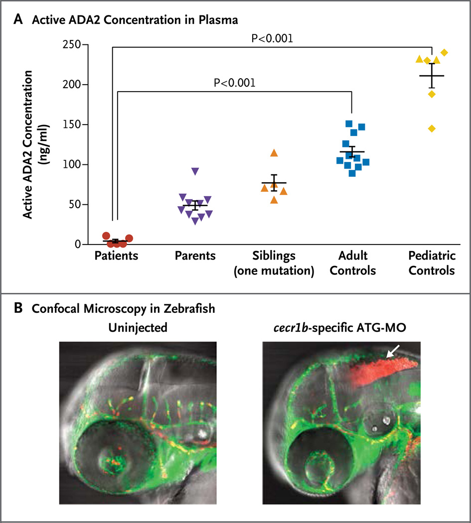
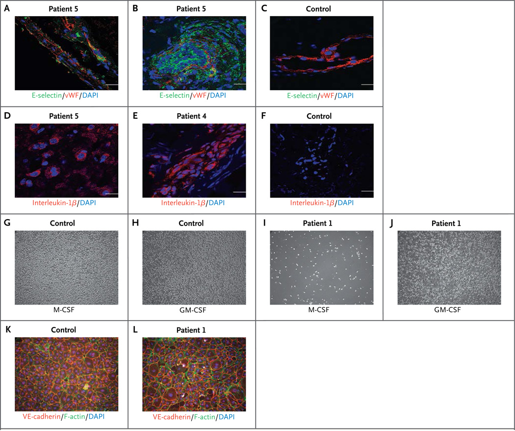
Results: All nine patients carried recessively inherited mutations in CECR1 (cat eye syndrome chromosome region, candidate 1), encoding adenosine deaminase 2 (ADA2), that were predicted to be deleterious; these mutations were rare or absent in healthy controls. Six patients were compound heterozygous for eight CECR1 mutations, whereas the three patients with polyarteritis nodosa or small-vessel vasculitis were homozygous for the p.Gly47Arg mutation. Patients had a marked reduction in the levels of ADA2 and ADA2-specific enzyme activity in the blood. Skin, liver, and brain biopsies revealed vasculopathic changes characterized by compromised endothelial integrity, endothelial cellular activation, and inflammation. Knockdown of a zebrafish ADA2 homologue caused intracranial hemorrhages and neutropenia - phenotypes that were prevented by coinjection with nonmutated (but not with mutated) human CECR1. Monocytes from patients induced damage in cocultured endothelial-cell layers.

Conclusions: Loss-of-function mutations in CECR1 were associated with a spectrum of vascular and inflammatory phenotypes, ranging from early-onset recurrent stroke to systemic vasculopathy or vasculitis. (Funded by the National Institutes of Health Intramural Research Programs and others.).


Figures

Figure 1
Figure 1. Clinical Findings and Pedigrees of Patients with Deficiency of Adenosine Deaminase 2 (ADA2)
Panel A shows livedo racemosa in Patient 1. Panels B and C show the results of a skin-punch biopsy of the left leg of Patient 4, which revealed vasculitis involving a medium-size blood vessel in the reticular dermis. Panel B shows a dense inflammatory infiltrate with fibrinoid necrosis, and Panel C, the complete occlusion of the vascular lumen (hematoxylin and eosin stain). Panels D through G show the results of brain imaging. Panels D and E show diffusion-weighted axial magnetic resonance images indicating acute small-vessel ischemia in the brain, with Panel D showing ischemia over the left thalamus and posterior limb of the left internal capsule in Patient 5 and Panel E showing ischemia in the left paramedian ventral midbrain in Patient 3; corresponding apparent diffusion coefficient maps are not shown. Panel F shows chronic ischemic changes on axial T1-weighted images in Patient 4 in the form of a small, oval cavitation (lacune) over the right thalamus that resulted from a prior small-vessel occlusion. Panel G shows hemorrhagic events in Patient 2, as detected on T2*-weighted (gradient echo) images. The localized areas of hypointensity over the right caudate head and posterior thalamus correspond to areas of focal accumulation of hemosiderin due to intraparenchymal bleeding. The arrows in Panels D through G point to the area of abnormality. Panel H shows multiple foci of petechial hemorrhages around small-size vessels in the white matter of the brain in Patient 5 (hematoxylin and eosin stain). Panel I shows the pedigrees of the nine affected patients. Patients 1 through 6 were compound heterozygous for eight mutations in CECR1 (cat eye syndrome chromosome region, candidate 1), and Patients 7, 8, and 9 were homozygous for one CECR1 mutation. Squares denote male family members, circles female family members, solid symbols affected family members, and open symbols unaffected family members. Shared CECR1 mutations are color-coded. NM denotes nonmutated.
Figure 2
Figure 2. Evidence of Loss-of-Function Mutations in ADA2
Panel A shows ADA2 activity in plasma samples from five patients with stroke (red circles), relatives who are clinically asymptomatic carriers of one mutation (purple triangles and orange triangles), healthy adult controls (blue squares), and healthy pediatric controls (yellow diamonds) (see the Supplementary Appendix). Two siblings (yellow triangles) who are not carriers of ADA2 mutations clustered appropriately with pediatric controls. Plasma specimens were titrated against recombinant ADA2 standards to quantitate the amount of nonmutant protein that would give equivalent activity. The horizontal lines represent mean values, and the I bars the standard error. Panel B shows confocal microscopy in zebrafish. Transgenic expression of enhanced green fluorescent protein under the fli1 promoter labels blood vessels green; expression of red fluorescent protein from discosoma species under the gata1 promoter labels erythrocytes red. Intracranial bleeding (arrow) is observed in embryos injected with cecr1b-specific morpholino oligonucleotides, targeting the translation initiation site (ATG-MO). Similar results were observed for embryos injected with the splice-blocking morpholinos targeting the proper splicing of exon 3.
Figure 3
Figure 3. Effect of ADA2 Deficiency in Patients
Cells stained with endothelial-cell activation marker E-selectin (green) are shown in a brain-biopsy sample from Patient 5 (Panel A) and in skin-biopsy samples from Patient 5 (Panel B) and a control donor (Panel C). E-selectin in endothelial cells (expressing von Willebrand factor [vWF; red]) indicates endothelial-cell activation (Panels A and B). E-selectin is absent in endothelial cells from a healthy control (Panel C). Nuclei are stained blue with 4′,6-diamidino-2-phenylindole (DAPI). Scale bars indicate 20 µm. Interleukin-1β immunostaining (red) is shown in a brain-biopsy sample from Patient 5 (Panel D) and in skin-biopsy samples from Patient 4 (Panel E) and a control donor (Panel F). Positive cells can be seen in the brain sample from Patient 5 (Panel D), and robust interleukin-1β staining is seen in a skin-biopsy sample from Patient 4 (Panel E). An M1 and M2 differentiation assay (Panels G through J) was performed in monocytes that had been isolated by means of negative selection from blood samples from Patient 1 and an age-matched control. Equal cell numbers were seeded; M2 macrophage differentiation was induced with 50 ng per milliliter of macrophage colony-stimulating factor (M-CSF), and M1 macrophage differentiation with 20 ng per milliliter of granulocyte–macrophage colony-stimulating factor (GM-CSF) for 10 days. Control monocytes attached and differentiated, showing macrophage-like morphologic features under both M-CSF and GM-CSF stimulation (Panels G and H). Very few attached and differentiated M2-like cells were observed in M-CSF–stimulated monocytes from Patient 1 (Panel I). However, M1-like cells were observed in GM-CSF–induced differentiation of monocytes from Patient 1 (Panel J) — similar to those seen in control cells. Human dermal microvascular endothelial cells were grown to confluence and cocultured with monocytes isolated from a control donor and from Patient 1 for 3 days. Nonadherent cells were removed, and the endothelial-cell layers were stained for the endothelial junction protein VE-cadherin (red), F-actin (green), and DAPI (blue). Endothelial cells cultured with healthy control monocytes (Panel K) show normal cell layers, whereas endothelial cells cocultured with monocytes isolated from Patient 1 show damaged, interrupted endothelial cell layers (Panel L).

Comment in

  • Vasculitis syndromes: New insights into the molecular basis of systemic vasculitis.
    Cid MC, Aróstegui JI. Cid MC, et al. Nat Rev Rheumatol. 2014 Jun;10(6):323-4. doi: 10.1038/nrrheum.2014.75. Epub 2014 May 13. Nat Rev Rheumatol. 2014. PMID: 24818673 No abstract available.
  • Mutant ADA2 in vasculopathies.
    Kastner DL, Zhou Q, Aksentijevich I. Kastner DL, et al. N Engl J Med. 2014 Jul 31;371(5):480-1. doi: 10.1056/NEJMc1405506. N Engl J Med. 2014. PMID: 25075844 No abstract available.
  • Mutant ADA2 in vasculopathies.
    van Montfrans J, Zavialov A, Zhou Q. van Montfrans J, et al. N Engl J Med. 2014 Jul 31;371(5):478. doi: 10.1056/NEJMc1405506. N Engl J Med. 2014. PMID: 25075845 No abstract available.
  • Mutant ADA2 in vasculopathies.
    Van Eyck L, Liston A, Meyts I. Van Eyck L, et al. N Engl J Med. 2014 Jul 31;371(5):478-9. doi: 10.1056/NEJMc1405506. N Engl J Med. 2014. PMID: 25075846 No abstract available.
  • Mutant ADA2 in vasculopathies.
    Bras J, Guerreiro R, Santo GC. Bras J, et al. N Engl J Med. 2014 Jul 31;371(5):478-80. doi: 10.1056/NEJMc1405506. N Engl J Med. 2014. PMID: 25075847 No abstract available.
  • Mutant ADA2 in vasculopathies.
    Van Eyck L, Liston A, Wouters C. Van Eyck L, et al. N Engl J Med. 2014 Jul 31;371(5):480. doi: 10.1056/NEJMc1405506. N Engl J Med. 2014. PMID: 25075848 No abstract available.
  • Inflammatory Web Catches Vessels.
    Chitalia V. Chitalia V. Sci Transl Med. 2014 Apr 16;6(232):232ec67. doi: 10.1126/scitranslmed.3009248. Sci Transl Med. 2014. PMID: 29977459 Free PMC article. No abstract available.

References

    1. Kastner DL, Aksentijevich I, Goldbach-Mansky R. Autoinflammatory disease reloaded: a clinical perspective. Cell. 2010;140:784–790. - PMC - PubMed
    1. Crow YJ, Rehwinkel J. Aicardi-Goutieres syndrome and related phenotypes: linking nucleic acid metabolism with autoimmunity. Hum Mol Genet. 2009;18(R2):R130–R136. - PMC - PubMed
    1. du Moulin M, Nürnberg P, Crow YJ, Rutsch F. Cerebral vasculopathy is a common feature in Aicardi-Goutieres syndrome associated with SAMHD1 mutations. Proc Natl Acad Sci U S A. 2011;108(26):E232–E233. - PMC - PubMed
    1. Lima LH, Schubert C, Ferrara DC, et al. Three major loci involved in age-related macular degeneration are also associated with polypoidal choroidal vasculopathy. Ophthalmology. 2010;117:1567–1570. - PMC - PubMed
    1. Kato GJ, Hebbel RP, Steinberg MH, Gladwin MT. Vasculopathy in sickle cell disease: biology, pathophysiology, genetics, translational medicine, and new research directions. Am J Hematol. 2009;84:618–625. - PMC - PubMed

Publication types

MeSH terms

Substances