. 2014;13(6):941-52.
doi: 10.4161/cc.27804. Epub 2014 Jan 16.

SmgGDS-558 regulates the cell cycle in pancreatic, non-small cell lung, and breast cancers

Affiliations

SmgGDS-558 regulates the cell cycle in pancreatic, non-small cell lung, and breast cancers

Nathan J Schuld et al. Cell Cycle. 2014.

Abstract

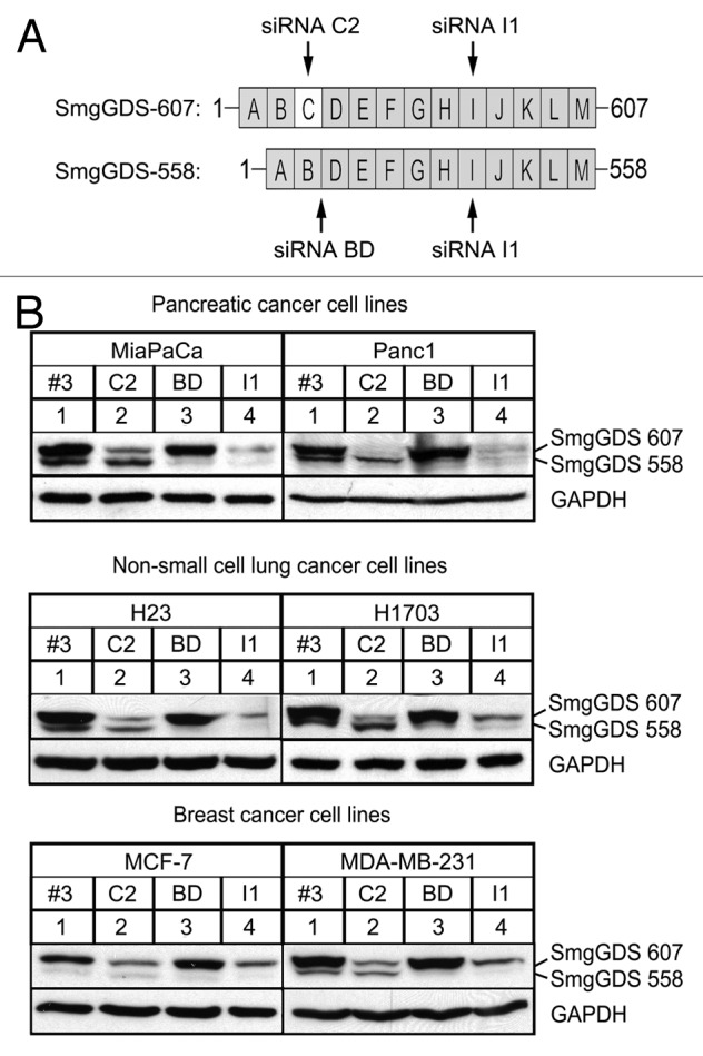
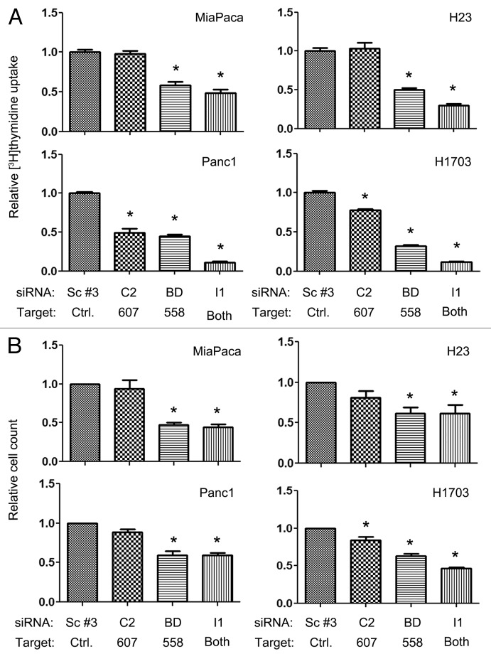
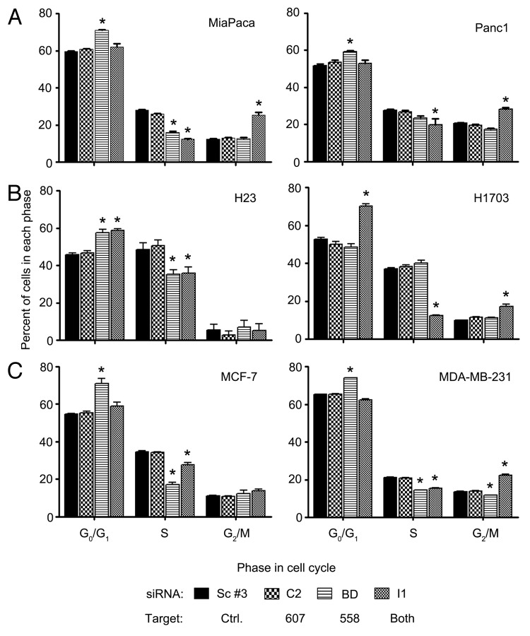
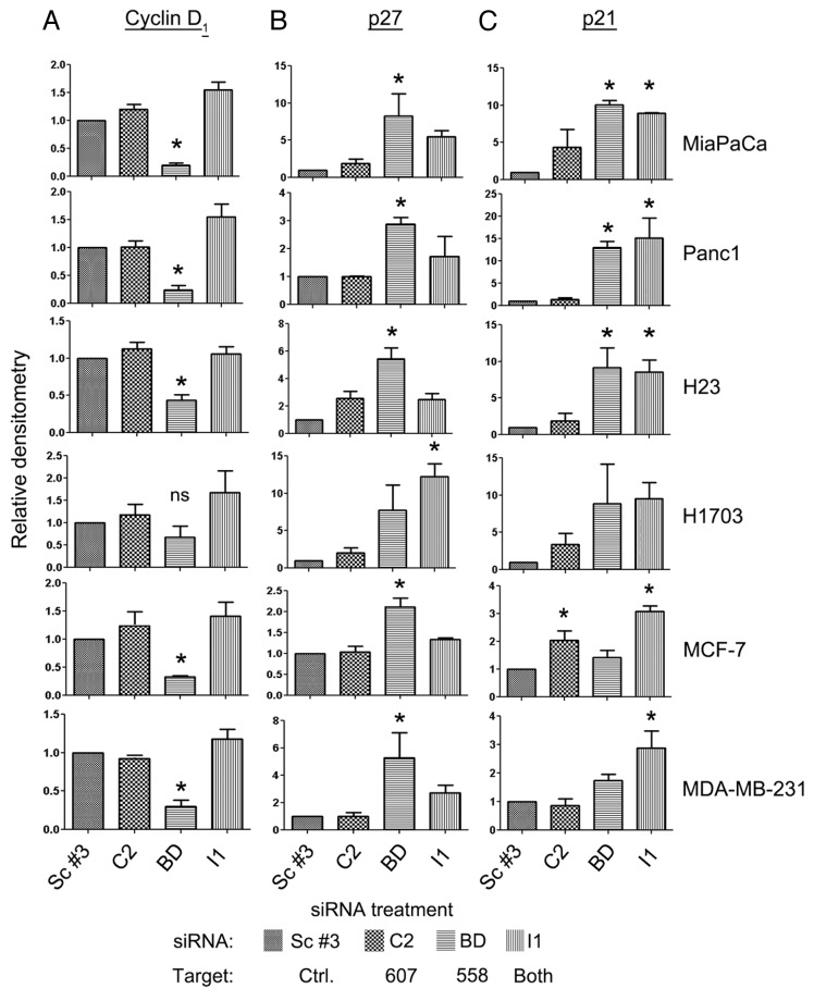
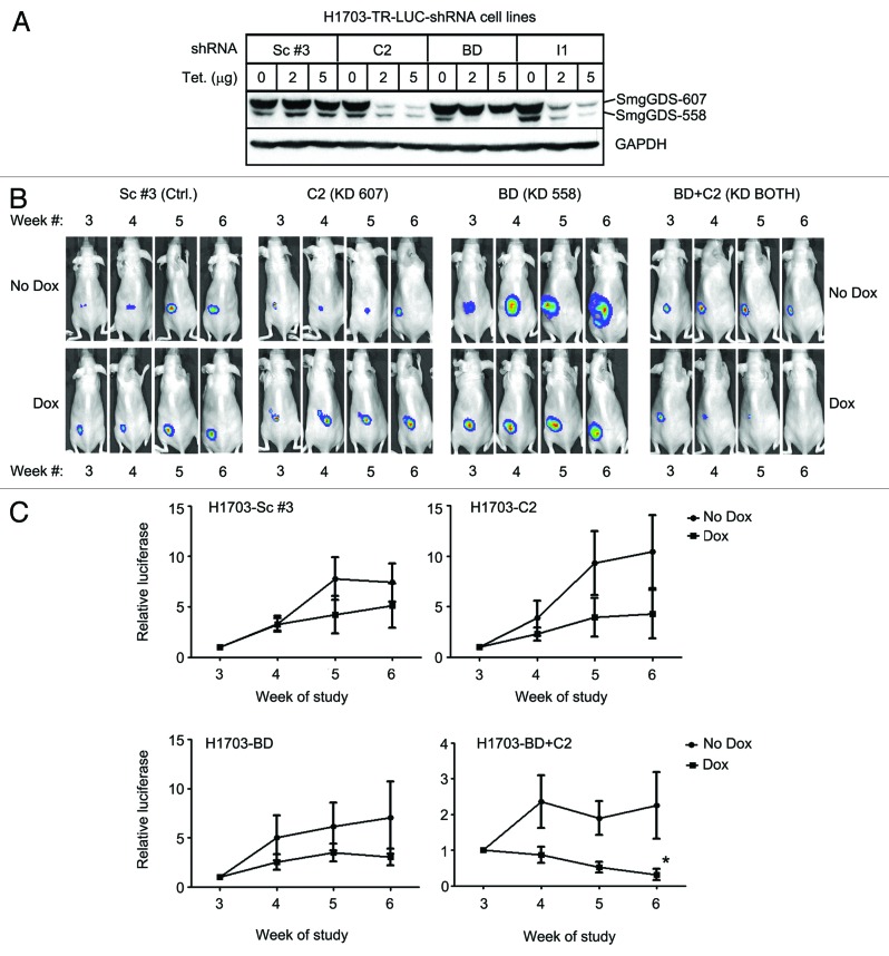
Oncogenic mutation or misregulation of small GTPases in the Ras and Rho families can promote unregulated cell cycle progression in cancer. Post-translational modification by prenylation of these GTPases allows them to signal at the cell membrane. Splice variants of SmgGDS, named SmgGDS-607 and SmgGDS-558, promote the prenylation and membrane trafficking of multiple Ras and Rho family members, which makes SmgGDS a potentially important regulator of the cell cycle. Surprisingly little is known about how SmgGDS-607 and SmgGDS-558 affect cell cycle-regulatory proteins in cancer, even though SmgGDS is overexpressed in multiple types of cancer. To examine the roles of SmgGDS splice variants in the cell cycle, we compared the effects of the RNAi-mediated depletion of SmgGDS-558 vs. SmgGDS-607 on cell cycle progression and the expression of cyclin D1, p27, and p21 in pancreatic, lung, and breast cancer cell lines. We show for the first time that SmgGDS promotes proliferation of pancreatic cancer cells, and we demonstrate that SmgGDS-558 plays a greater role than SmgGDS-607 in cell cycle progression as well as promoting cyclin D1 and suppressing p27 expression in multiple types of cancer. Silencing both splice variants of SmgGDS in the cancer cell lines produces an alternative signaling profile compared with silencing SmgGDS-558 alone. We also show that loss of both SmgGDS-607 and SmgGDS-558 simultaneously decreases tumorigenesis of NCI-H1703 non-small cell lung carcinoma (NSCLC) xenografts in mice. These findings indicate that SmgGDS promotes cell cycle progression in multiple types of cancer, making SmgGDS a valuable target for cancer therapeutics.

Keywords: GTPase; RNAi; Rap1GDS1; SmgGDS; breast cancer; cell cycle; lung cancer; mouse tumorigenesis; pancreatic cancer; proliferation.


Figures

None
Figure 1. SmgGDS-607 and SmgGDS-558 are expressed in pancreatic cancer, NSCLC, and breast cancer, and can be silenced utilizing SmgGDS siRNA. (A) SmgGDS splice variants 607 and 558 are comprised of 13 or 12 armadillo repeats (ARMs), respectively. SmgGDS-607 has an ARM C, which is spliced out of SmgGDS-558. The following siRNAs were utilized in this study: the siRNA C2 targets only SmgGDS-607, whereas siRNA BD targets only SmgGDS-558. The siRNA I1 targets both splice variants and the non-targeting control siRNA is labeled as Scramble #3. (B) Pancreatic cancer cells lines MiaPaCa and Panc1 (top), non-small cell lung cancer cell lines H23 and H1703 (middle), and breast cancer cell lines MCF-7 and MDA-MB-231 (bottom) were transfected with 25 nM of the indicated siRNAs, lysed after 72 h (h), and subjected to ECL-western blotting using antibodies to SmgGDS and GAPDH. Results are representative of 3 independent experiments.
None
Figure 2. Cell proliferation is diminished more by silencing SmgGDS-558 than by silencing SmgGDS-607, and silencing both splice variants together enhances this effect. The indicated cell lines were assayed for cell proliferation utilizing [3H]-thymidine uptake (A) or relative cell count (B) 72 h after transfecting the cells with 25 nM of the indicated siRNAs. The values are normalized to cells transfected with non-targeting scramble #3 siRNA. Results are the mean ± SE from 3 or more independent experiments conducted with either quadruplicate or sextuplicate (A) samples in each experiment. (*P < 0.01 by one-way ANOVA with Dunnett post-hoc multiple comparisons test.)
None
Figure 3. Silencing SmgGDS-558 alone or both SmgGDS-558 and SmgGDS-607 together causes a G1 or G2 cell cycle arrest. Pancreatic (A), NSCLC (B), or breast (C) cancer cell lines were transfected with 25 nM of the indicated siRNA and changes in cell cycle progression were determined by staining the cells with propidium iodide 72 h post-transfection, followed by fluorescence-activated cell sorter analysis. Results are the mean ± SE from 3 or more independent experiments. The symbol above a column indicates a statistical comparison of progression through each phase of the cell cycle by the indicated cells vs. the control cells transfected with Scramble #3 siRNA. (*P < 0.05)
None
Figure 4. Expression of Cyclin D1, p27, and p21 is altered in pancreatic, NSCLC, and breast cancer cells after silencing SmgGDS splice variants. Cyclin D1 (A), p27Kip1 (p27) (B), and p21/WAF1 (p21) (C) protein expression was analyzed in the indicated pancreatic, NSCLC, and breast cancer cell lines 72 h after transfection of the indicated SmgGDS siRNA. An equal number of live cells were lysed in SDS sample buffer and immunoblotted using antibodies to the cell cycle proteins. Densitometric analysis of the immunoblotted proteins was conducted by comparing the O.D. of the indicated cell cycle protein normalized to the O.D. of GAPDH for each treatment, and expressed as a percent of normalized protein expression in the cells treated with the control Scramble #3 siRNA. Results are the mean ± SE from 3 independent experiments. (ns, not significant; *P < 0.05 by one-way ANOVA with Dunnett post-hoc multiple comparisons test.)
None
Figure 5. Silencing both SmgGDS-607 and SmgGDS-558 together diminishes tumorigenesis by H1703 xenografts in mice. (A) H1703-LUC-TR cell lines stably expressing the indicated inducible shRNAs were cultured with increasing amounts of tetracycline (0, 2, or 5 μg) daily for 72 h. The cells were then lysed and subjected to ECL-western blotting using antibodies to SmgGDS and GAPDH. Results are representative of 3 independent experiments. (BandC) H1703-LUC-TR cell lines stably expressing the indicated inducible shRNAs were injected into the flank of SHO mice. At week 3, doxycycline (dox) was introduced via the diet of half of the animals in each group. Representative mouse images (B) are shown for each cell line (KD = knockdown of the indicated splice variant). The graphs (C) represent relative weekly growth of tumors: H1703-Sc #3 (top left; n = 7 mice without dox and n = 6 mice with dox), H1703-C2 (top right; n = 6 mice without dox and n = 6 mice with dox), H1703-BD (bottom left; n = 6 mice without dox and n = 6 mice with dox), and H1703-BD+C2 (bottom right; n = 6 mice without dox and n = 7 mice with dox.) Values are normalized to luminescence obtained at week 3 for each mouse and are the mean ± SE assessed by 2-way ANOVA with secondary Bonferroni multiple comparisons test (*P < 0.05).

Similar articles

Cited by

References

    1. Siegel R, Naishadham D, Jemal A. Cancer statistics, 2013. CA Cancer J Clin. 2013;63:11–30. doi: 10.3322/caac.21166. - DOI - PubMed
    1. Garcea G, Neal CP, Pattenden CJ, Steward WP, Berry DP. Molecular prognostic markers in pancreatic cancer: a systematic review. Eur J Cancer. 2005;41:2213–36. doi: 10.1016/j.ejca.2005.04.044. - DOI - PubMed
    1. Kusama T, Mukai M, Iwasaki T, Tatsuta M, Matsumoto Y, Akedo H, Nakamura H. Inhibition of epidermal growth factor-induced RhoA translocation and invasion of human pancreatic cancer cells by 3-hydroxy-3-methylglutaryl-coenzyme a reductase inhibitors. Cancer Res. 2001;61:4885–91. - PubMed
    1. Taniuchi K, Nakagawa H, Hosokawa M, Nakamura T, Eguchi H, Ohigashi H, Ishikawa O, Katagiri T, Nakamura Y. Overexpressed P-cadherin/CDH3 promotes motility of pancreatic cancer cells by interacting with p120ctn and activating rho-family GTPases. Cancer Res. 2005;65:3092–9. - PubMed
    1. Liu Y, Wang Y, Zhang Y, Miao Y, Zhao Y, Zhang PX, Jiang GY, Zhang JY, Han Y, Lin XY, et al. Abnormal expression of p120-catenin, E-cadherin, and small GTPases is significantly associated with malignant phenotype of human lung cancer. Lung Cancer. 2009;63:375–82. doi: 10.1016/j.lungcan.2008.12.012. - DOI - PubMed

Publication types

MeSH terms