Review
. 2014;13(6):898-909.
doi: 10.4161/cc.28255. Epub 2014 Feb 17.

Senescence-accelerated OXYS rats: a model of age-related cognitive decline with relevance to abnormalities in Alzheimer disease

Affiliations
Review

Senescence-accelerated OXYS rats: a model of age-related cognitive decline with relevance to abnormalities in Alzheimer disease

Natalia A Stefanova et al. Cell Cycle. 2014.

Abstract

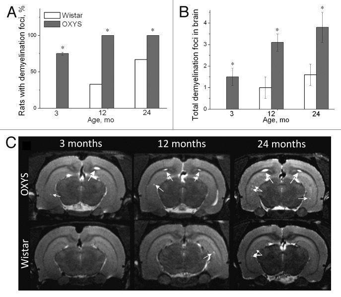
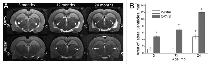
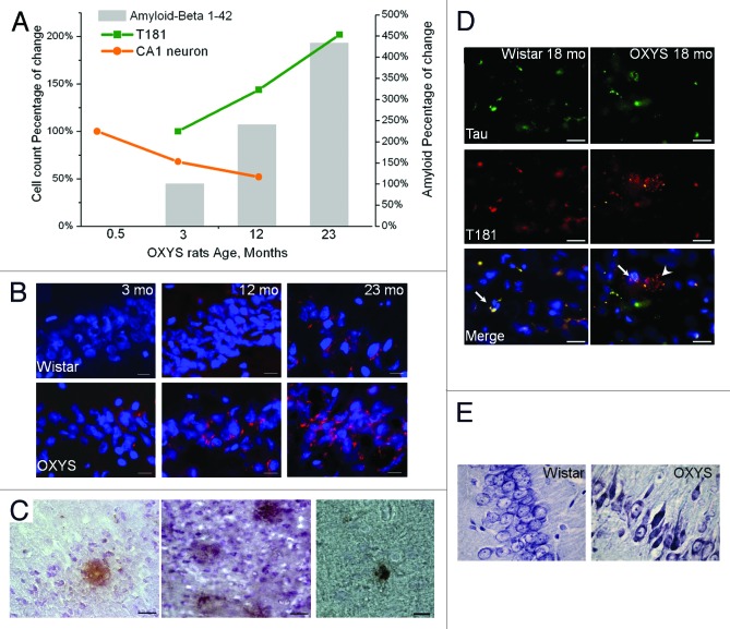
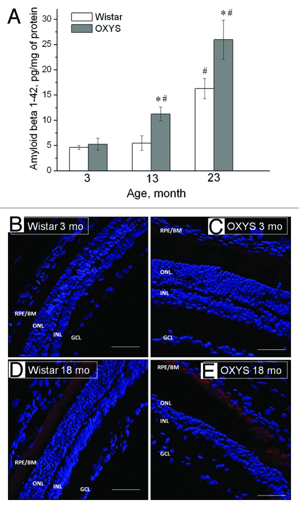
Senescence-accelerated OXYS rats are an experimental model of accelerated aging that was established from Wistar stock via selection for susceptibility to cataractogenic effects of a galactose-rich diet and via subsequent inbreeding of highly susceptible rats. Currently, we have the 102nd generation of OXYS rats with spontaneously developing cataract and accelerated senescence syndrome, which means early development of a phenotype similar to human geriatric disorders, including accelerated brain aging. In recent years, our group found strong evidence that OXYS rats are a promising model for studies of the mechanisms of brain aging and neurodegenerative processes similar to those seen in Alzheimer disease (AD). The manifestation of behavioral alterations and learning and memory deficits develop since the fourth week of age, i.e., simultaneously with first signs of neurodegeneration detectable on magnetic resonance imaging and under a light microscope. In addition, impaired long-term potentiation has been demonstrated in OXYS rats by the age of 3 months. With age, neurodegenerative changes in the brain of OXYS rats become amplified. We have shown that this deterioration happens against the background of overproduction of amyloid precursor protein (AβPP), accumulation of β-amyloid (Aβ), and hyperphosphorylation of the tau protein in the hippocampus and cortex. The development of AMD-like retinopathy in OXYS rats is also accompanied by increased accumulation of Aβ in the retina. These published data suggest that the OXYS strain may serve as a spontaneous rat model of AD-like pathology and could help to decipher the pathogenesis of AD.

Keywords: Alzheimer disease; brain aging; senescence-accelerated OXYS rats.


Figures

None
Figure 1. Quantification of neuronal populations in the hippocampal CA3 region of OXYS rats. (A) No cell loss in the CA3 region was detected at 0.5 mo; furthermore, 5-mo-old OXYS rats showed increased neuronal populations (P < 0.05); however, 15-mo-old OXYS rats showed a cell loss (P < 0.05) when compared with age-matched Wistar rats (control). *A statistically significant difference between the strains of the same age; #significant age-related differences compared with the previous age within the strain. (B) The neurodegenerative changes occur in the CA3 region of the hippocampus of OXYS rats: the percentage of dead or damaged neurons is significantly larger than that in Wistar rats at 5 and 15 mo of age. 1 (light gray) corresponds intact neurons, 2 (black) to dead neurons, and 3 (gray) to damaged neurons. Adapted from (Maksimova et al., in press).
None
Figure 2. T2-weighed MRI images of demyelinating foci in 3-, 12-, and 24-mo-old Wistar and OXYS rats. (A) The presence of foci of demyelization in 3-, 12-, and 24-mo-old Wistar and OXYS rats (% animals). (B) OXYS rats have a greater number of demyelinating foci compared with Wistar rats. (C) Axial slices of the brain of 3-, 12-, and 24-mo-old Wistar and OXYS rats. The arrows point to foci of demyelization. The data are shown as mean ± SEM. Legend: *P < 0.05 for differences between the strains. Adapted from reference .
None
Figure 3. T2-weighed MRI images of lateral ventricles in 3-, 12-, and 24-mo-old Wistar and OXYS rats. (A) Axial slices of the brain of 3-, 12-, and 24-mo-old Wistar and OXYS rats. Increases in the size of lateral ventricles in OXYS rats are visible (the arrows). (B) In 3-, 12-, and 24-mo-old OXYS rats there was an enlargement of the lateral ventricles compared with Wistar rats. The data are shown as mean ± SEM. Legend: *P < 0.05 for differences between the strains. Adapted from reference .
None
Figure 4. Time course of progression of AD-like pathology, as a percentage of either 0.5- or 3-mo-old OXYS rats in a group. (A) Rats exhibited a 47% loss of neurons in the CA1 region of the hippocampus between 0.5 and 12 mo of age. In addition, a 102% increase in the phospho-tau T181 level occurred between 3 and 23 mo of age. Total Aβ expression increased by 333% between 3 and 23 mo of age. (B) Aβ1–42 level is increased in the hippocampus of 12- and 23-mo-old but not 3-mo-old OXYS rats compared with disease-free (control) Wistar rats. Staining for Aβ1–42 (red). DAPI staining (blue) corresponds to cellular nuclei. Scale bars: 20 μm. (C) Photomicrographs demonstrate the Aβ deposits in the brain of OXYS rats detected by MOAB-2, clone 6C3. Scale bars: 50 μm. (D) Immunostaining for Tau (green) and phospho-tau T181 (red) in the cortex of 18-mo-old OXYS and Wistar rats. DAPI staining (blue) corresponds to cellular nuclei. The arrow shows colocalization of Tau and phospho-tau T181 in OXYS and Wistar rats, whereas the arrowhead shows phospho-tau T181 in OXYS rats. Scale bars: 20 μm. (E) The percentages of dead or damaged neurons in the CA1 region of the hippocampus in 15-mo-old OXYS rats are significantly greater compared with Wistar rats. Neurons were stained with cresyl violet. Adapted from reference and Maksimova et al.
None
Figure 5.1–42 level is increased in the retina of OXYS rats compared with disease-free (control) Wistar rats. (A) Levels of Aβ1–42 of OXYS and Wistar rats at the age of 3, 13, and 23 mo (ELISA). *A statistically significant difference between the strains of the same age; #significant age-related differences compared with the previous age within the strain. (BE) Confocal immunofluorescent images depict a weak Aβ (red) signal detected in a 3-mo-old Wistar rat (B), a stronger signal in a 3-mo-old OXYS rat (C), and strong staining in 18-mo-old rats of both strains (D and E). Cell nuclei are stained with DAPI (blue). The scale bar is 50 μm. RPE/BrM, retinal pigment epithelium/Bruch membrane; ONL, outer nuclear layer; INL, inner nuclear layer; GCL, ganglion cell layer. Adapted from reference .

Similar articles

Cited by

References

    1. Feng Y, Wang X. Antioxidant therapies for Alzheimer’s disease. Oxid Med Cell Longev. 2012;2012:472932. doi: 10.1155/2012/472932. - DOI - PMC - PubMed
    1. Querfurth HW, LaFerla FM. Alzheimer’s disease. N Engl J Med. 2010;362:329–44. doi: 10.1056/NEJMra0909142. - DOI - PubMed
    1. Morley JE, Armbrecht HJ, Farr SA, Kumar VB. The senescence accelerated mouse (SAMP8) as a model for oxidative stress and Alzheimer’s disease. Biochim Biophys Acta. 2012;1822:650–6. doi: 10.1016/j.bbadis.2011.11.015. - DOI - PubMed
    1. Teich AF, Arancio O. Is the amyloid hypothesis of Alzheimer’s disease therapeutically relevant? Biochem J. 2012;446:165–77. doi: 10.1042/BJ20120653. - DOI - PMC - PubMed
    1. Liao FF, Wang R, Park EA. Repression of Alzheimer’s beta-secretase. Aging (Albany NY) 2013;5:789–90. - PMC - PubMed

Publication types

MeSH terms

Substances