. 2014;13(6):992-1005.
doi: 10.4161/cc.27880. Epub 2014 Jan 22.

Distinctive effects of the cellular inhibitor of apoptosis protein c-IAP2 through stabilization by XIAP in glioblastoma multiforme cells

Affiliations

Distinctive effects of the cellular inhibitor of apoptosis protein c-IAP2 through stabilization by XIAP in glioblastoma multiforme cells

Wensheng Yang et al. Cell Cycle. 2014.

Abstract

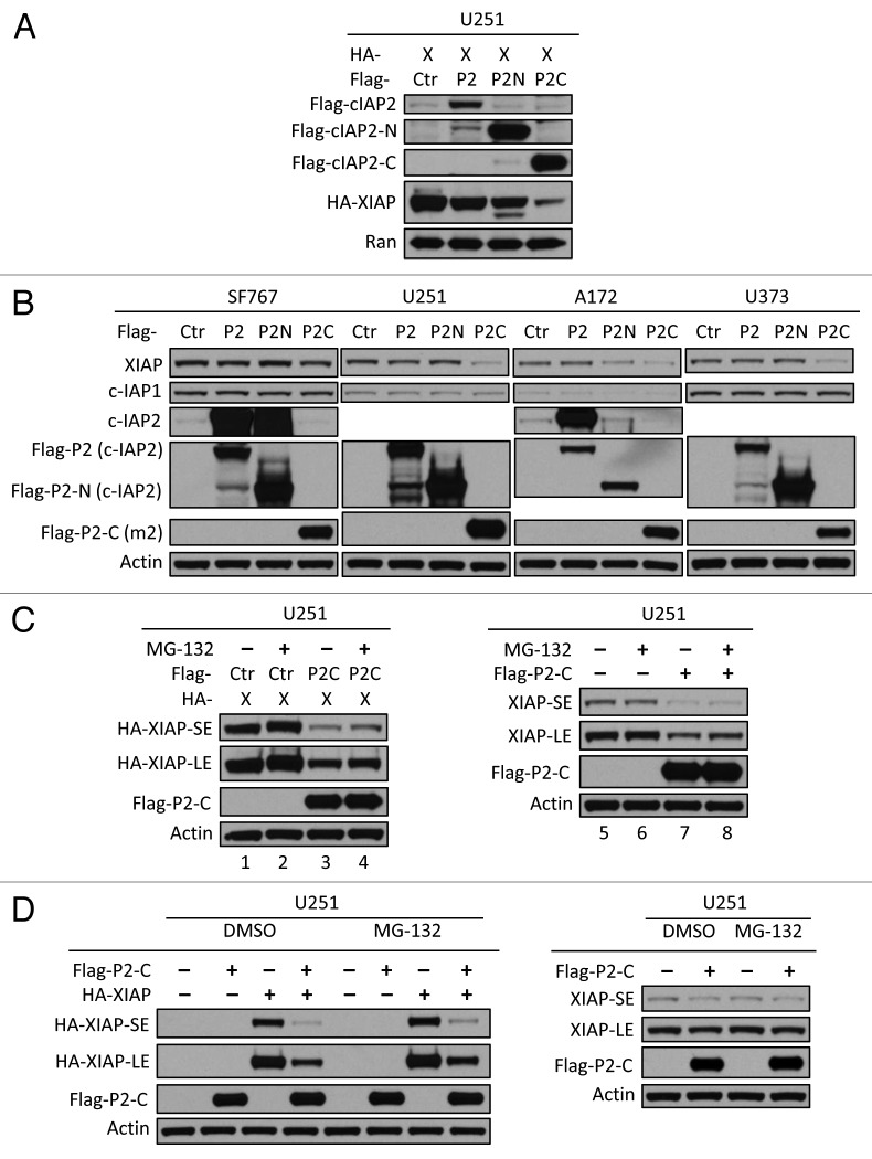
Inhibitor of apoptosis proteins (IAPs) are extensively involved in NFκB signaling pathways. Regulation of c-IAP2 turnover by other proteins was investigated in glioblastoma multiforme (GBM) cells in the present study. When overexpressed, X-linked IAP (XIAP) enhanced expression of ectopic c-IAP2, but not c-IAP1, and endogenous c-IAP2 levels were reduced once XIAP expression was silenced. TNFα stimulation substantially increased c-IAP2 expression, and this upregulation was impaired by suppression of XIAP. Similarly, when XIAP was limiting due to severe hypoxic conditions, c-IAP2 levels were downregulated. These data together indicate that XIAP is an important regulator responsible for stabilization of c-IAP2 levels under different conditions. Protein interactions occur through binding of BIR2 and BIR3 domains of c-IAP2 with the RING finger of XIAP. XIAP inhibition of c-IAP2 auto-degradation was dependent on this physical interaction, and it was independent of XIAP E3 ligase activity. Global c-IAP2 ubiquitination was not affected by XIAP, although c-IAP2 levels were significantly increased. A CARD-RING-containing fragment of c-IAP2 was found to target XIAP for proteasome-independent degradation, but it was unable to sensitize GBM cells to chemo-reagents. The XIAP-stabilized c-IAP2 was found to enhance IκB-α phosphorylation on serines 32 and 36, and to antagonize XIAP-induced increase in mature Smac and Bcl10. Taken together, our data identify a distinctive role of c-IAP2 as stabilizer of XIAP, which is likely involved in regulation of NFκB activation and apoptosis in GBM cells.

Keywords: Bcl10; IκB-α; TNFα; XIAP; anoxia; auto-degradation; c-IAP2; glioblastoma multiforme cell lines; mature Smac.


Figures

None
Figure 1. XIAP enhances the expression level of c-IAP2. (A) Four glioblastoma multiforme (GBM) cell lines (A172, SF767, U251, and U373) and normal human astrocytes were assessed by western blot analysis using the indicated antibodies to determine the steady-state levels of XIAP, c-IAP1, and c-IAP2. Ran was used as a loading control. (B) Plasmid DNAs of Flag-c-IAP1 (P1), or Flag-c-IAP2 (P2) with or without Flag-XIAP (X), each at 6 µg, were transfected into U251 or U373 cells. The cells were harvested after 40−48 h, and then protein levels were measured by western blot analysis using anti-Flag m2 antibody and other antibodies as indicated. Arrow indicates a non-specific band. (C) Plasmid myc-c-IAP2 (P2) with control (C), or Flag-XIAP (X) plasmids was transfected into U251 or U373 cells, and the cells were lysed after 40−48 h and protein levels were assessed by immunoblotting as indicated. (D) Five micrograms of plasmid Myc-c-IAP2 were transfected into U251 or U373 cells with an increasing amount of Flag-XIAP plasmids (from 1 to 5 µg), followed by immunoblotting to examine overexpression of ectopic proteins. Beta-actin was used as a loading control (1 min, 30 s, and 10 s refer to the film exposure times). (E) A172 and SF767 cells were transfected with siRNA to silence XIAP expression. After 72 h, c-IAP2, XIAP, and Ran expression was determined by immunoblotting. In addition, HCT116 cells with wild-type or null XIAP were directly analyzed by immunoblotting (middle panel), or first transfected with control (C) or myc-c-IAP2 (P2) plasmids (right panel) and then after 24 h assessed by immunoblotting (wt, wild-type; ko, knockout; SE, short-time exposure, LE, long-time exposure).
None
Figure 2A–C. XIAP is required for the stability of c-IAP2 in different stimulatory settings. (A) GBM cells were treated with 40 ng/ml of TNFα for 22 h, and then endogenous c-IAP1, c-IAP2, and XIAP expression was assessed by immunoblotting as indicated. (B) U251 or U373 cells were transfected with control siRNA or XIAP siRNA (C, control; X, XIAP siRNA). Approximately 24 h post-transfection cells were treated with TNFα (40 ng/ml) or control for 24 h followed by immunoblotting for c-IAP2, XIAP, and Ran expression as indicated. (C) HCT116 or MEF cells (XIAP wild-type and XIAP knocked out) were treated with 20 ng/ml or 40ng/ml of TNFα for 2 h or 2–8 h as indicated. The figure shows representative immunoblots. The experiment was performed 3 times with similar results. The integrated optical density of each band was quantitated and is shown below the immunoblots. c-IAP2 values were normalized against the corresponding RAN band and expressed as arbitrary units (AU).
None
Figure 2D. (D) HCT cells (XIAP wild-type or knocked out) were treated under anoxia for 1 h, 3 h, or 5 h. The figure shows representative immunoblots. The experiment was performed 3 times with similar results. The integrated optical density of each band was quantitated and is shown below the immunoblots. c-IAP2 values were normalized against the corresponding RAN band and expressed as arbitrary units (AU).
None
Figure 3. XIAP interacts with c-IAP2. (A) U373 cells were transfected with Flag-XIAP (X), together with Myc-c-IAP2 (P2) or control empty vector (C) plasmids. Two days later, cells were lysed and immunoprecipitated (IPed) with anti-Flag m2 antibodies. The co-IPed proteins were assessed by immunoblotting as indicated. (B) Four human GBM cell lines, A172, SF767, U251, and U373, were lysed with IP buffer, and each cell lysate was IPed with rabbit anti-human c-IAP2 antibody or mouse anti-human XIAP antibody, and co-IPed proteins were analyzed by immunoblotting as shown. (C) SF767 cell lysates were IP-ed with rabbit anti-c-IAP2 antibody (P2), or normal rabbit IgG (RIgG), or rabbit anti-antibodies (Luc), and assessed for the specificity of co-IPed XIAP.
None
Figure 4. Mapping the regions responsible for interaction between c-IAP2 and XIAP. (A) Schematic representation of family-associated domains of c-IAP2 and its deletion mutants used for mapping. (B) Plasmid expressing Flag-tagged c-IAP2 N terminus (aa 1–441, Flag-P2N) or Flag-tagged c-IAP2 C terminus (aa 442–604, Flag-P2C) was transfected to SF767 cells, and then after 24 h the cells were lysed in IP buffer. The cell lysates were incubated with anti-Flag M2-agarose affinity gel and then proteins in the co-IP were assessed by western blot analysis using anti-human XIAP antibody or anti-Flag, as indicated. (C) SF767 cells were transfected with 6 µg of Flag-c-IAP2-N (N), Flag-c-IAP2-ΔBIR1 (ΔB1), or Flag-c-IAP2-BIR2/3 (B2/3) plasmid DNA. After 24 h, the cells were harvested in IP buffer and subjected to IP with anti-Flag M2-agarose affinity gel. Co-IPed proteins were examined by immunoblotting, as indicated. (D) Schematic representation of family-associated domains of XIAP and its deletion mutants. (E) U251 cells were transfected with 5 µg of Flag-c-IAP2 (P2) and 5 µg of HA-XIAP (X), HA-XIAP-ΔRING (ΔR), or HA-XIAP-ΔBIR (ΔB) plasmid DNA. After 24 h, IP was conducted using mouse anti-HA antibody and then proteins in co-IP were assessed in immunoblots, as indicated. (SE, short exposure; LE, long exposure; PC, picture corrections).
None
Figure 7. C-terminal fragment of c-IAP2 targets endogenous IAPs for degradation. (A) Flag-c-IAP2 (P2), Flag-c-IAP2-N (P2N), or Flag-c-IAP2-C (P2C) and HA-XIAP plasmid DNAs were transfected into U251 cells. After 24 h, the cells were lysed and then assessed in immunoblots, as indicated. (B) In 4 GBM cell lines, 5 µg of Flag-c-IAP2 (P2), Flag-c-IAP2-N (P2N), or Flag-c-IAP2-C (P2C) plasmid DNAs were transfected, and 24 h later cells were lysed and endogenous IAP degradation was analyzed by immunoblotting. (C and D) Flag-c-IAP2-C plasmid DNA with or without HA-XIAP was transfected into U251 cells, and 24 h later the cells were treated with 10 µM MG-132 for 8 h in (C) or 40 µM MG-132 for 4 h in (D). The expression level of HA-XIAP or endogenous XIAP was determined, as indicated. (SE, short exposure; LE, long exposure).
None
Figure 6. Binding to c-IAP2 is sufficient for XIAP stabilization. (A) U251 or U373 cells were transfected with 5 µg of Flag-c-IAP2 (or HA-c-IAP2) and 5 µg of control, HA-XIAP, or HA-XIAP-H467A plasmid DNA, and then, approximately 40 h later, cell lysates were assessed in immunoblots, as indicated. (B) U373 cells were transfected with 5 µg of HA-c-IAP2 and 5 µg of control, HA-XIAP, or HA-XIAP-ΔRING plasmid DNA, and then 44 h later cell lysates were assessed by immunoblotting as indicated. (PC, picture correction). (C) U251 cells were transfected with 5 µg of Flag-c-IAP2, Flag-c-IAP2-ΔBIR1, or Flag-c-IAP2-N plasmid DNA with or without 5 µg of Flag-XIAP as indicated. (C, control; P2, c-IAP2; ΔB1, c-IAP2-ΔBIR1; N, c-IAP2-N). Approximately 40–44 h post-transfection, the cells were lysed in sample buffer and then protein expression was measured by western blot analysis, as indicated.
None
Figure 5. XIAP inhibits c-IAP2 auto-degradation. (A) SF767 cells were transfected with Flag-XIAP (X) and HA-tagged c-IAP2 (P2), c-IAP2-H574A (574), or control (Ctr) plasmid DNA. About 48 h later, cell lysates were assessed in immunoblots, as indicated. (B) U251 cells were transfected with Flag-c-IAP2 (P2) or Flag-c-IAP2-N (P2N) plasmid DNA; in addition, SF767 cells were transfected with HA-c-IAP2-H574A (574) or control (Ctr) plasmid. After 20 h, the cells were treated with 10 µM MG-132. After 8 h, cell lysates were assessed in immunoblots, as indicated. (C) U251 cells were transfected with Flag-XIAP or myc-c-IAP2 plasmid DNA or both, and 40 h later they were treated with 10 µM MG-132. After 8 h, ubiquitinated c-IAP2 in cell lysates was examined in immunoblots. (D) U251 cells were transfected with 5 µg of Flag-XIAP and myc-c-IAP2 plasmid DNA and then 20 h later they were treated with 10 µM MG-132. After 8 h, the cells were lysed in IP buffer and cell lysates were incubated with anti-Flag m2 affinity gel. The myc-c-IAP2 in co-IP was determined.
None
Figure 8. XIAP shapes the c-IAP2 E3 ligase activity onto its targets. (A) U251 cells were transfected with control (abbreviated for C), or c-IAP2 (myc- or Flag-tagged, P2), or Flag-XIAP (X) or both (P2/X) along with Flag-IκB-α followed by immunoblotting assessment 20–40 h post-transfection as indicated. (B) U251 cells were transfected with 5 µg of plasmid control, or Flag-c-IAP1, or myc-c-IAP2, or Flag-XIAP along with 0.05 µg of Δ55Smac-HA followed by immunoblotting assessment 24 h post-transfection. In addition, 4 cell lines were transfected, respectively, with 5 µg of plasmid control or Flag-XIAP with 0.1 µg of Δ55Smac-HA and assessed by immunoblotting 24 h later as indicated. (C) A172 cells were transfected with 0.1 µg of Δ55Smac-HA in addition to myc-c-IAP2, or Flag-XIAP, or both at 5 µg each (or as indicated otherwise) and analyzed by immunoblotting 19–22 h post-transfection. (D) U251 cells were transfected with 0.05–0.1 µg of Δ55Smac-HA and myc-c-IAP2 or Flag-XIAP or both with 5 µg each (or as indicated otherwise) and analyzed by immunoblotting 19–24 h post-transfection. (E) U373 or A172 cells were transfected with 5 µg of plasmid control or Flag-/myc- tagged c-IAP2 (FP2/mP2) and assessed by immunoblotting 24 h post-transfection as indicated. (F) A172 cells were transfected with plasmid control, or myc-c-IAP2, or Flag-XIAP, or both followed by immunoblotting analysis 40 h post-transfection as indicated.
None
Figure 9. Possible mechanisms by which c-IAP2 is regulated. TNFα signaling through TNFR1-mediated canonical NFκB activation transcriptionally upregulates c-IAP2 in a XIAP-dependent manner, while XIAP-enhanced c-IAP2 facilitates the phosphorylation of IκB-α leading to its subsequent proteasomal degradation. In addition, anoxic conditions improve c-IAP2 levels by increased XIAP and likely active p53 signaling. Finally, XIAP in human GBM cell lines stabilizes mature Smac (ΔSmac) and Bcl10 antagonized by c-IAP2, implying distinctive activities of c-IAP2 among IAPs (black arrow, upregulation; solid orange arrow, documented transcriptional activation; square dot orange arrow, speculative activation; rightward-turned capital letter T, inhibition).

Similar articles

Cited by

References

    1. Crook NE, Clem RJ, Miller LK. An apoptosis-inhibiting baculovirus gene with a zinc finger-like motif. J Virol. 1993;67:2168–74. - PMC - PubMed
    1. Srinivasula SM, Ashwell JD. IAPs: what’s in a name? Mol Cell. 2008;30:123–35. doi: 10.1016/j.molcel.2008.03.008. - DOI - PMC - PubMed
    1. Gyrd-Hansen M, Meier P. IAPs: from caspase inhibitors to modulators of NF-kappaB, inflammation and cancer. Nat Rev Cancer. 2010;10:561–74. doi: 10.1038/nrc2889. - DOI - PubMed
    1. Yang Y, Fang S, Jensen JP, Weissman AM, Ashwell JD. Ubiquitin protein ligase activity of IAPs and their degradation in proteasomes in response to apoptotic stimuli. Science. 2000;288:874–7. doi: 10.1126/science.288.5467.874. - DOI - PubMed
    1. Hu S, Yang X. Cellular inhibitor of apoptosis 1 and 2 are ubiquitin ligases for the apoptosis inducer Smac/DIABLO. J Biol Chem. 2003;278:10055–60. doi: 10.1074/jbc.M207197200. - DOI - PubMed

Publication types

MeSH terms

Substances