. 2014;13(6):1041-8.
doi: 10.4161/cc.28128. Epub 2014 Feb 7.

The NAD+ synthesizing enzyme nicotinamide mononucleotide adenylyltransferase 2 (NMNAT-2) is a p53 downstream target

Affiliations

The NAD+ synthesizing enzyme nicotinamide mononucleotide adenylyltransferase 2 (NMNAT-2) is a p53 downstream target

Lu-Zhe Pan et al. Cell Cycle. 2014.

Abstract

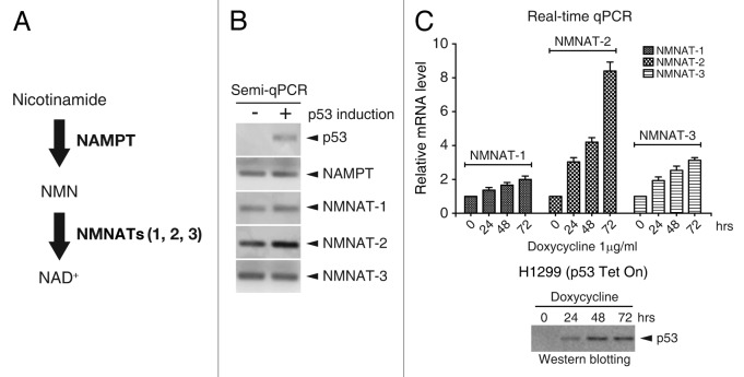
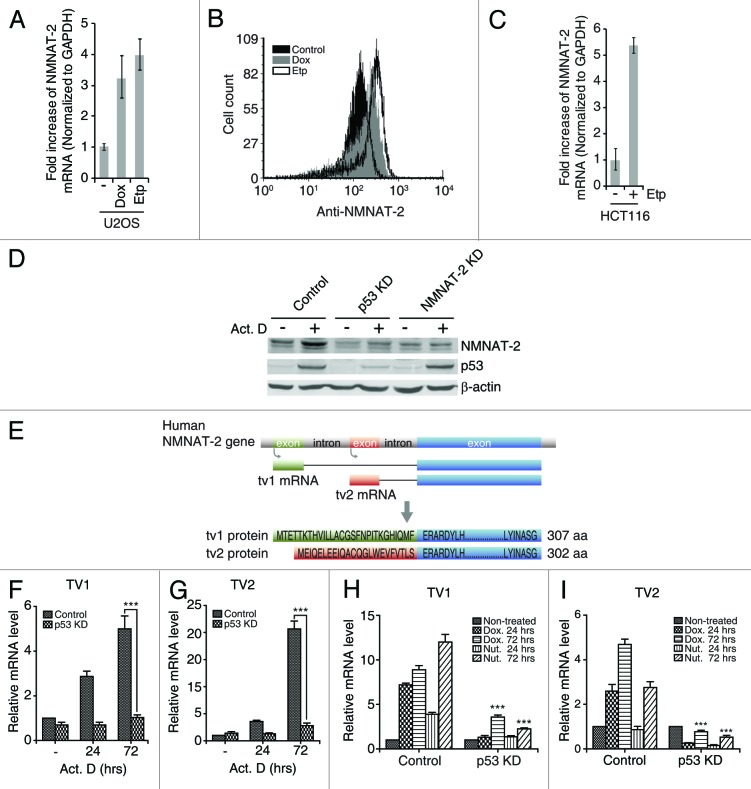
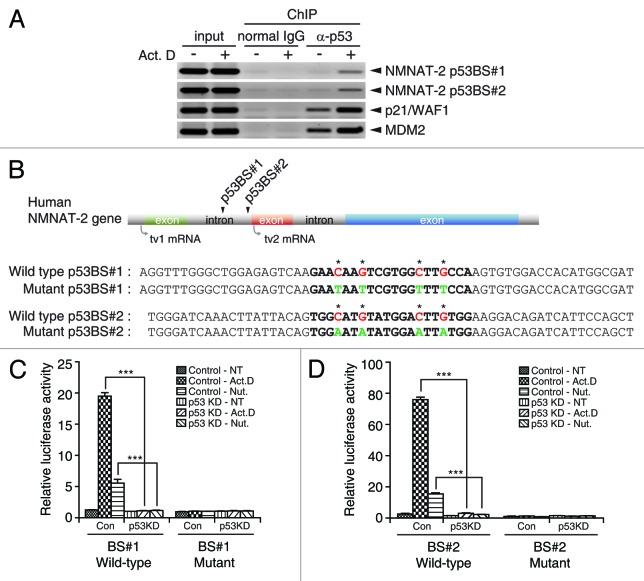
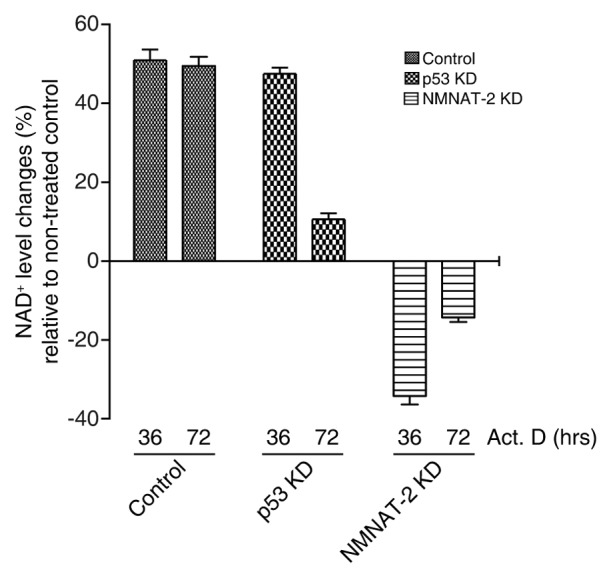
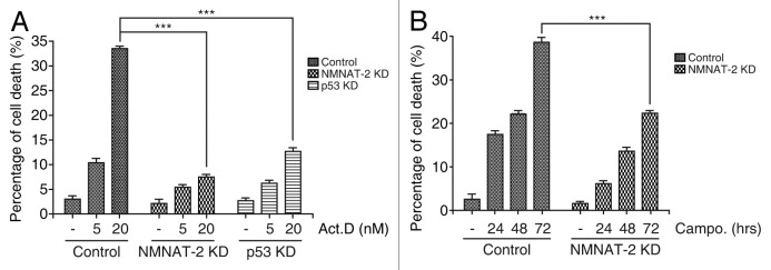
NAD(+) metabolism plays key roles not only in energy production but also in diverse cellular physiology. Aberrant NAD(+) metabolism is considered a hallmark of cancer. Recently, the tumor suppressor p53, a major player in cancer signaling pathways, has been implicated as an important regulator of cellular metabolism. This notion led us to examine whether p53 can regulate NAD(+) biosynthesis in the cell. Our search resulted in the identification of nicotinamide mononucleotide adenylyltransferase 2 (NMNAT-2), a NAD(+) synthetase, as a novel downstream target gene of p53. We show that NMNAT-2 expression is induced upon DNA damage in a p53-dependent manner. Two putative p53 binding sites were identified within the human NMNAT-2 gene, and both were found to be functional in a p53-dependent manner. Furthermore, knockdown of NMNAT-2 significantly reduces cellular NAD(+) levels and protects cells from p53-dependent cell death upon DNA damage, suggesting an important functional role of NMNAT-2 in p53-mediated signaling. Our demonstration that p53 modulates cellular NAD(+) synthesis is congruent with p53's emerging role as a key regulator of metabolism and related cell fate.

Keywords: NAD+ biosynthesis; NMNAT-2; apoptosis; p53.


Figures

None
Figure 1. p53 induces NMNAT-2 expression. (A) Schematic diagram of NAD+ biosynthesis via the salvage pathway. (B and C) Cellular mRNA levels of NAMPT, NMNAT-1, NMNAT-2, and NMNAT-3 in response to p53 induction were determined by semi- (B) or real-time qRT-PCR (C). Relative mRNA levels were normalized to those from controls cells (no doxycycline). p53 expression in H1299-p53-Tet On cells was induced by 1 μg/ml of doxycycline (C, bottom panel). Data represent mean values from 3 independent experiments, with error bars showing SEM.
None
Figure 2. NMNAT-2 induction by DNA damage is p53-dependent. (A–C) NMNAT-2 expression was measured in U2OS and HCT116 cells treated with doxorubicin (Dox, 200 ng/ml) or etoposide (Etp, 4 μM). mRNA level of NMNAT-2 was measured using real-time qRT-PCR after 72 h (A and C). Cell-associated NMNAT-2 protein levels were compared by flow cytometry with intracellular staining after 48 h (B). (D) U2OS cells (control, p53-KD and NMNAT-2-KD) were treated with actinomycin D (Act. D, 20 nM) for 48 h. Protein levels of p53 and NMNAT-2 were compared. Actin was used as loading control. (E) Schematic illustration of the human NMNAT-2 gene and its encoded products. Transcription start sites and protein sequences of N-terminal of NMNAT-2 transcription variant 1 (tv1) and 2 (tv2) are shown. (F–I) U2OS cells (control or p53-KD) were treated with actinomycin D (20 nM), doxorubicin (200 ng/ml), or nutlin-3 (Nut, 10 μM) for the indicated times. Fold-increase of mRNA in U2OS p53-KD cells were compared with that of U2OS control cells. Data represent mean values from 3 independent experiments, with error bars showing SEM: *P < 0.05; **P < 0.01; ***P < 0.0001.
None
Figure 3. NMNAT-2 is a direct target of p53. (A) p53 binds to 2 putative binding sites. U2OS cells treated with actinomycin D (20 nM) were subjected to chromatin immunoprecipitation (ChIP) analysis. Chromatin fragments co-immunoprecipitated with p53 protein were amplified using specific primers spanning individual p53REs. p53REs in the p21WAF1/CIP1 and mdm2 gene were used as positive controls. Mouse monoclonal anti-p53 antibody was used for p53 pull-down and mouse normal IgG for negative control. Results are representative of 2 independent experiments. Arrowheads indicate the amplified REs. (B) Location of p53 binding sites (BS) upstream of NMNAT-2 gene was depicted. Sequences of wild-type and mutant p53 BS#1 and BS#2 are shown. Consensus sequences of p53-response elements (REs) are designated in bold lettering with nucleotide substitutions indicated by asterisks. Wild-type nucleotides of p53BS#1 and #2 highlighted in red were substituted with indicated nucleotides in green as depicted in mutant-type. (C and D) p53REs identified in human NMNAT-2 gene are transcriptionally active. Firefly luciferase reporter plasmids carrying individual p53BS#1 and #2 (wild-type or mutant) were co-transfected with Renilla luciferase (for normalization) into U2OS cells (control or p53-KD). Cells were treated with actinomycin D (10 nM) or nutlin-3 (10 nM) for 48 h. Luciferase activities of each reporter were normalized to those from non-treated U2OS control cells. Data are presented as mean values with error bars showing SEM from 3 independent experiments. *P < 0.05; **P < 0.01; ***P < 0.0001.
None
Figure 4. NMNAT-2 regulates cellular NAD+ level. U2OS cells (control, p53-KD, and NMNAT-2-KD) were treated with actinomycin D (20 nM) for the indicated times, and cellular NAD+ level was then measured. Data presented are mean values from 3 independent experiments with error bars showing SEM.
None
Figure 5. NMNAT-2 is required for p53-dependent cell death. Knocking down NMNAT-2 protects U2OS cells from DNA damage-induced cell death. (A) U2OS cells (control, NMNAT-2-KD and p53-KD) were treated with actinomycin D (5 or 20 nM) for 48 h. The percentage of dead cells was determined by sub-G1 DNA content analysis using flow cytometry. (B) Cells were treated with camptothecin (0.5 μM) for 24 h. Data presented as mean values from 3 independent experiments with error bars showing SEM. Cell death in NMNAT-2-KD or p53-KD cells was compared with that of U2OS control cells. *P < 0.05; **P < 0.01; ***P < 0.0001.

Comment in

  • p53, DNA damage, and NAD+ homeostasis.
    Pfister NT, Yoh KE, Prives C. Pfister NT, et al. Cell Cycle. 2014;13(11):1661-2. doi: 10.4161/cc.29151. Epub 2014 May 8. Cell Cycle. 2014. PMID: 24810733 Free PMC article. No abstract available.

References

    1. Di Stefano G, Manerba M, Vettraino M. NAD metabolism and functions: a common therapeutic target for neoplastic, metabolic and neurodegenerative diseases. Curr Top Med Chem. 2013;13:2918–29. - PubMed
    1. Imai S, Armstrong CM, Kaeberlein M, Guarente L. Transcriptional silencing and longevity protein Sir2 is an NAD-dependent histone deacetylase. Nature. 2000;403:795–800. doi: 10.1038/35001622. - DOI - PubMed
    1. Ahn BH, Kim HS, Song S, Lee IH, Liu J, Vassilopoulos A, Deng CX, Finkel T. A role for the mitochondrial deacetylase Sirt3 in regulating energy homeostasis. Proc Natl Acad Sci U S A. 2008;105:14447–52. doi: 10.1073/pnas.0803790105. - DOI - PMC - PubMed
    1. Hirschey MD, Shimazu T, Goetzman E, Jing E, Schwer B, Lombard DB, Grueter CA, Harris C, Biddinger S, Ilkayeva OR, et al. SIRT3 regulates mitochondrial fatty-acid oxidation by reversible enzyme deacetylation. Nature. 2010;464:121–5. doi: 10.1038/nature08778. - DOI - PMC - PubMed
    1. van der Horst A, Tertoolen LG, de Vries-Smits LM, Frye RA, Medema RH, Burgering BM, van der HA FOXO4 is acetylated upon peroxide stress and deacetylated by the longevity protein hSir2(SIRT1) J Biol Chem. 2004;279:28873–9. doi: 10.1074/jbc.M401138200. - DOI - PubMed

Publication types

Substances

LinkOut - more resources