. 2014 Feb 19;281(1780):20133368.
doi: 10.1098/rspb.2013.3368. Print 2014 Apr 7.

Generation of mastitis resistance in cows by targeting human lysozyme gene to β-casein locus using zinc-finger nucleases

Affiliations

Generation of mastitis resistance in cows by targeting human lysozyme gene to β-casein locus using zinc-finger nucleases

Xu Liu et al. Proc Biol Sci. .

Abstract

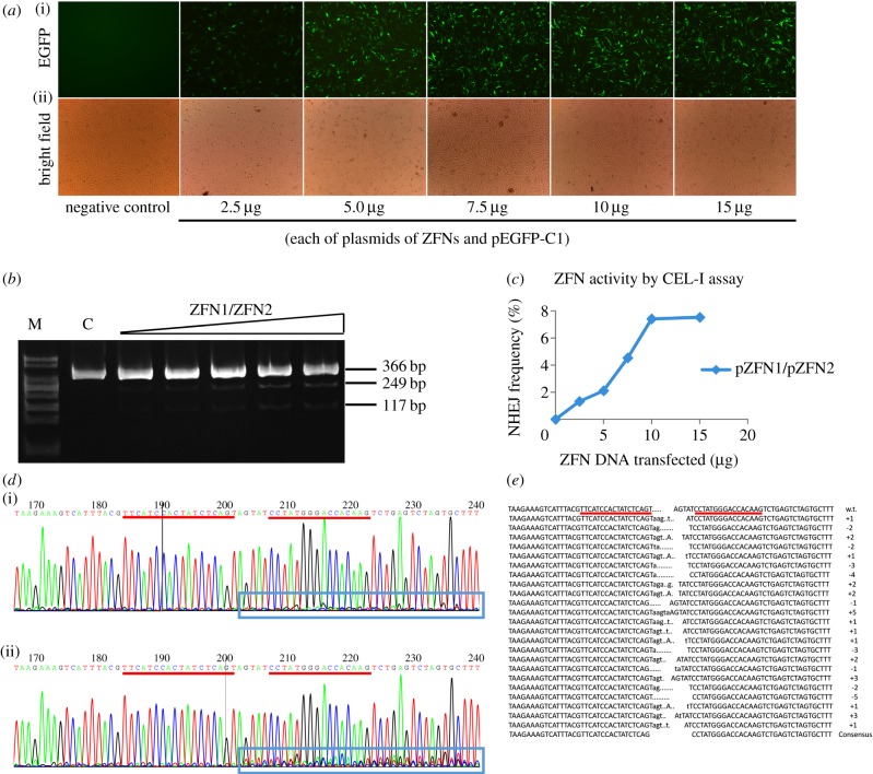
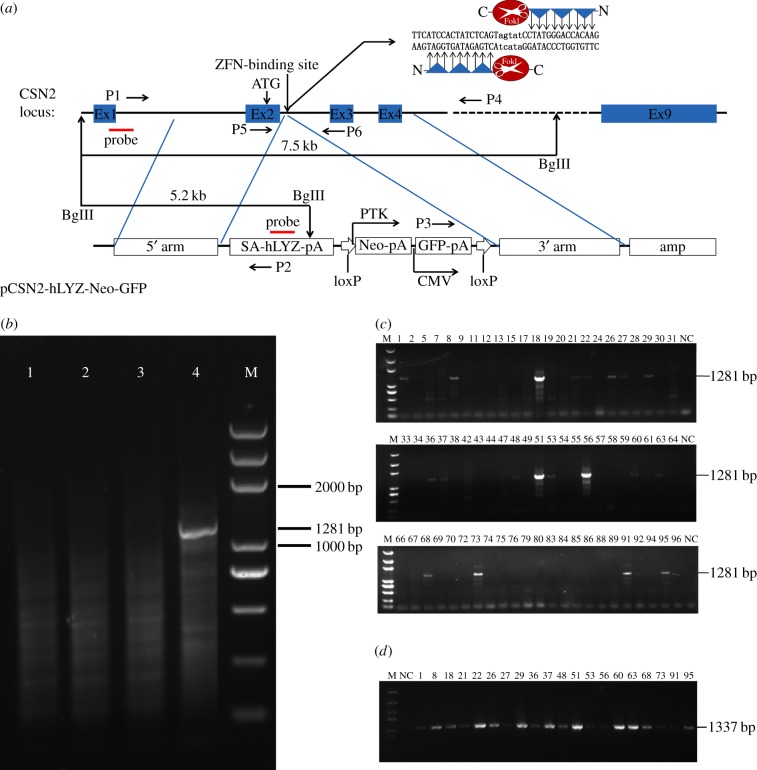
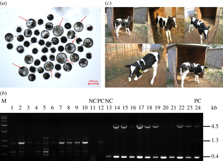
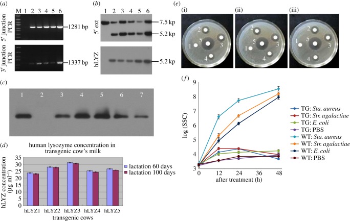
Mastitis costs the dairy industry billions of dollars annually and is the most consequential disease of dairy cattle. Transgenic cows secreting an antimicrobial peptide demonstrated resistance to mastitis. The combination of somatic cell gene targeting and nuclear transfer provides a powerful method to produce transgenic animals. Recent studies found that a precisely placed double-strand break induced by engineered zinc-finger nucleases (ZFNs) stimulated the integration of exogenous DNA stretches into a pre-determined genomic location, resulting in high-efficiency site-specific gene addition. Here, we used ZFNs to target human lysozyme (hLYZ) gene to bovine β-casein locus, resulting in hLYZ knock-in of approximately 1% of ZFN-treated bovine fetal fibroblasts (BFFs). Gene-targeted fibroblast cell clones were screened by junction PCR amplification and Southern blot analysis. Gene-targeted BFFs were used in somatic cell nuclear transfer. In vitro assays demonstrated that the milk secreted by transgenic cows had the ability to kill Staphylococcus aureus. We report the production of cloned cows carrying human lysozyme gene knock-in β-casein locus using ZFNs. Our findings open a unique avenue for the creation of transgenic cows from genetic engineering by providing a viable tool for enhancing resistance to disease and improving the health and welfare of livestock.

Keywords: gene targeting; mastitis; nuclear transfer; zinc-finger nucleases; β-casein locus.


Figures

Figure 1.
Figure 1.
Validation of ZFNs for the CSN2 locus in bovine fibroblasts. (a) (i) EGFP direct fluorescence microscopy and (ii) phase-contrast microscopy of bovine fibroblasts that were mock transfected (mock) and of bovine fibroblasts that were transfected with increasing amounts of ZFN expression constructs pZFN1/pZFN2 mixed with plasmid pEGFP-C1. Micrographs were acquired 72 h post-transfection. Numerals below the various columns correspond to the amounts of the ZFN expression plasmids and of the plasmid pEGFP-C1. Original magnification, 100×. (b) The frequency of allelic mutation in each pool of treated cells was determined using the CEL-I assay (gel). Bands migrating at 366, 249 and 117 bp represent the parent amplicon and the two CEL-I digestion products, respectively. The bands were quantitated by ethidium bromide staining and densitometry to determine the frequency of NHEJ. (c) The frequency of NHEJ is plotted against ZFN dosage. (d) Sequence analysis showed that the presence of multiple peaks after the targeted site in the sequencing curves clearly distinguishes (i) non-targeted cells from (ii) mutants. PCR products corresponding to the targeted site were sequenced directly. (e) Sequence alignment revealed distinct ZFN-induced insertions and deletions within the target region of CSN2.
Figure 2.
Figure 2.
Targeting of CSN2 in bovine fibroblasts. (a) Schematic overview depicting the targeting strategy for the CSN2 locus. Blue boxes, exons of CSN2; vertical arrow, the translational initiation signal (ATG) of the β-casein; bold vertical arrow, genomic site cut by the ZFN pair; horizontal arrows, primers used for junction PCR; red bars indicate the probe used for Southern blot analysis. The predicted size of Southern hybridization bands with BglII digestion, for both the endogenous CSN2 locus and the CSN2 targeted locus, is indicated. The positions and orientations of the loxP sites are indicated by thick white arrows. Shown above is a schematic of the donor plasmid design. The donor plasmid was created to correspond to the cleavage location of the ZFN pairs and carried a roughly 700 bp region of homology to the CSN2 sequence around the cleavage site. SA, splice acceptor sequence; hLYZ, human lysozyme gene sequence; pA, polyadenylation signals; Neo, neomycin resistance gene; GFP, enhanced green fluorescent protein gene; PTK, protein tyrosine kinase promoter; CMV, human cytomegalovirus immediate early promoter; Amp, ampicillin resistance gene. The inset at the upper right is a cartoon of ZFNs binding at a specific genomic site (upper case), which leads to the dimerization of the FokI nuclease domains. (b) PCR-based measurements of ZFN-driven exogenous gene integration into the CSN2 locus in bovine fibroblasts. Cells were left untransfected (lane 1, for negative control) or were transfected with an expression cassette for ZFNs that induce a DSB at intron 2 of CSN2 (lane 2), and donor plasmids carrying a foreign gene flanked by 700 bp homology arms, in the absence (lane 3) and presence (lane 4) of the CSN2 ZFNs. Genomic DNA was extracted 72 h later. The CSN2 locus was amplified by 30 cycles of PCR in the presence of radiolabelled dNTPs by using primers P1 and P2 specific for the CSN2 locus and the foreign gene, respectively. (c) 5′ junction PCR analysis carried out on chromosomal DNA of parental bovine fibroblasts (NC) and of clones derived from bovine fibroblasts co-transfected with ZFNs expression constructs pZFN1/pZFN2 and gene-targeting vector pCSN2-hLYZ-Neo-GFP. HR-mediated transgene insertion should yield 1281 bp PCR products using primers P1 and P2, which are specific for the CSN2 locus and the exogenous gene, respectively. (d) 3′ junction PCR analysis carried out on genomic DNA from 5′ junction PCR-positive colonies using primers P3 and P4 to amplify the 1337 bp right-hand junction between endogenous and exogenous DNA.
Figure 3.
Figure 3.
Bovine gene-targeted cloned embryos developed in vitro. (a) The bovine reconstructed embryos were cultured in vitro. Grade I blastocysts are indicated as red arrows. (b) Ten nuclear cloned embryos were used for individual-embryo PCR assays to rule out mixed colonies, as many targeted colonies also contained non-targeted cells. Lanes 1–12, 5′ junction PCR analysis using primers P1 and P2; lane 11, non-transgene cloning embryo used for negative control; lane 12, gene-targeted cell colony used for positive control. Lanes 13–24, long-range PCR analysis using primers P5 and P6; lane 13, non-transgene cloning embryo used for negative control; lane 24, gene-targeted cell colony used for positive control. (c) The gene-targeted cows at one month postpartum (born July 2010).
Figure 4.
Figure 4.
Analysis of knock-in cows. (a) PCR analysis carried out on chromosomal DNA of non-transgenic cow (lane 1) and of five gene-targeted calves lived for more than one month (lanes 2–6). 5′ junction PCR should yield 1281 bp PCR products using primers P1 and P2 (upper), 3′ junction PCR should yield 1337 bp PCR products using primers P3 and P4 (lower). The positions and sizes of specific PCR products are indicated at the right. (b) Southern blot analysis of the CSN2 knock-in calves. Lane 1 contains normal cow DNA digested with BglII as a negative control. Lanes 2–6 are BglII-digested genomic DNA from five gene-targeted calves. Using the external probe 5′ of the genome homology region, the CSN2 knock-in calves showed two bands: a 7.5 kb band from the endogenous CSN2 allele and a 5.2 kb band characteristic of the insertion (upper). The 5.2 kb band was also detected with an hLYZ probe (lower). (c) Western blot analysis of milk probed with antibody against recombinant human lysozyme: lane 1 contains bacterially derived recombinant human lysozyme in saline (50 ng); lane 2 contains 1 µl milk from non-transgenic cow; lanes 3–7 each contains 1 µl milk from gene-targeted cows. (d) Human lysozyme concentrations were measured by ELISA during the first lactation of transgenic cows. (e) Bacterial plate assay for bacteriolytic activity. Lytic zones were developed on the lawn of (i) Sta. aureus, (ii) E. coli and (iii) Str. agalactiae 12 h after sample application: 10 µl milk from non-transgenic cow (1) and 10 µl milk from gene-targeted cows (2, 3, 4) and 10 µl (500 ng ml−1) recombinant human lysozyme (5). (f) Somatic cell concentration in milk of transgenic (n = 5) and non-transgenic (n = 5) cows to intra-mammary infusion of 80 c.f.u. of Sta. aureus, E. coli and Str. agalactiae. The vertical coordinate is the log value of the somatic cell count.

References

    1. Klug A. 2010. The discovery of zinc fingers and their applications in gene regulation and genome manipulation. Annu. Rev. Biochem. 79, 213–231. (10.1146/annurev-biochem-010909-095056) - DOI - PubMed
    1. Urnov FD, Rebar EJ, Holmes MC, Zhang HS, Gregory PD. 2010. Genome editing with engineered zinc finger nucleases. Nat. Rev. Genet. 11, 636–646. (10.1038/nrg2842) - DOI - PubMed
    1. Lombardo A, et al. 2007. Gene editing in human stem cells using zinc finger nucleases and integrase-defective lentiviral vector delivery. Nat. Biotechnol. 25, 1298–1306. (10.1038/nbt1353) - DOI - PubMed
    1. Urnov FD, et al. 2005. Highly efficient endogenous human gene correction using designed zinc-finger nucleases. Nature 435, 646–651. (10.1038/nature03556) - DOI - PubMed
    1. Kim YG, Cha J, Chandrasegaran S. 1996. Hybrid restriction enzymes: zinc finger fusions to Fok I cleavage domain. Proc. Natl Acad. Sci. USA 93, 1156–1160. (10.1073/pnas.93.3.1156) - DOI - PMC - PubMed

Publication types

MeSH terms