Review
. 2014 Apr;5(100):42-9.
doi: 10.1016/j.coviro.2014.01.008. Epub 2014 Feb 16.

A cool hybrid approach to the herpesvirus 'life' cycle

Affiliations
Review

A cool hybrid approach to the herpesvirus 'life' cycle

Tzviya Zeev-Ben-Mordehai et al. Curr Opin Virol. 2014 Apr.

Abstract

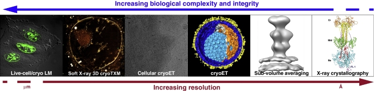
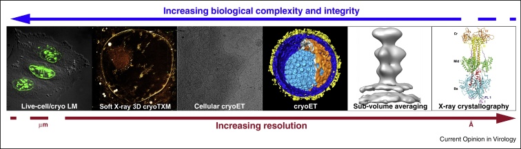
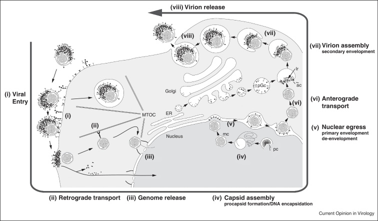
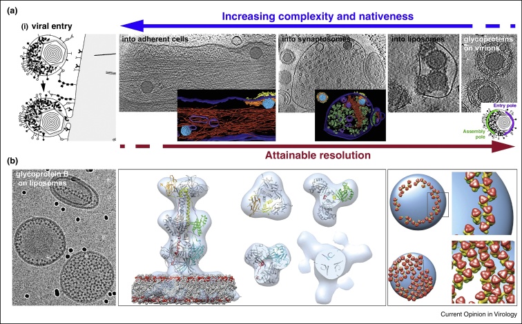
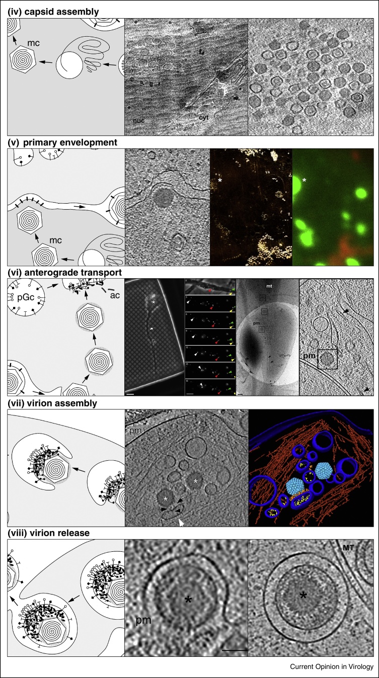
Electron cryo tomography (cryoET) is an ideal technique to study virus-host interactions at molecular resolution. Imaging of biological specimens in a frozen-hydrated state assures a close to native environment. Various virus-host cell interactions have been analysed in this way, with the herpesvirus 'life' cycle being the most comprehensively studied. The data obtained were further integrated with fluorescence and soft X-ray cryo microscopy data applied on experimental systems covering a wide range of biological complexity. This hybrid approach combines dynamic with static imaging and spans a resolution range from micrometres to angstroms. Along selected aspects of the herpesvirus replication cycle, we describe dedicated combinations of approaches and how subsequent data integration enables insights towards a functional understanding of the underlying processes.


Figures

None
Graphical abstract
Figure 1
Figure 1
The spectrum of techniques applied to study the herpesvirus ‘life’ cycle. An integrated approach combining high-resolution structure determination methods and correlative light, soft X-ray cryo and electron cryo microscopy allows looking at dynamic processes at different resolution and complexity and ultimately leads to a better perception of those processes. LM, light microscopy; cryoTXM, transmission X-ray cryo microscopy; cryoET, electron cryo tomography.
Figure 2
Figure 2
Illustration of the herpesvirus ‘life’ cycle. Virus infection starts with cell entry that involves the fusion of the viral envelope with that of the host, either directly at the plasma membrane or in an endosome leading to release of the capsid into the cytosol (i) [40]. The capsid is then transported retrogradely to the nucleus along microtubules (ii) [58]. The viral genome is released into the nucleus through the nuclear pore (iii) [59]. Transcription of viral genes and genome replication occur in the nucleus, as well as procapsid (pc) formation and DNA encapsidation (iv) [30]. Capsids exit the nucleus by a primary envelopment and de-envelopment mechanism (v) [43]. Capsids are then transported anterogradely to the point of virion assembly (vi). Virion assembly, including secondary envelopment and tegumentation, occurs close to the cell surface by budding into cellular vesicles originating from the Golgi that contain the viral glycoproteins on the lumenal side and accrete tegument proteins on the cytosolic (vii). Virions are released from the cell by fusion of the cellular vesicles with the plasma membrane (viii).
Figure 3
Figure 3
Dedicated experimental systems used for the study of HSV1 entry. (a) Reducing biological complexity from the most native system, virus entry into intact host cells [24], to gradually less complex subsystems, that is, virus entry into synaptosomes [24] or liposomes, and study of glycoproteins on the viral surface. (b) HSV1 glycoprotein B (gB), a key component of the fusion machinery, bound to liposomes used as display platform for direct visualization of fusion protein interaction with its target membrane. Sub-volume averaging was used to reconstruct the gB–lipid bilayer complex (middle panel, light grey) [42]. The EM reconstruction together with fitting of the gB crystal structure revealed the mode of interaction to the membrane. Lateral interaction of gB induced protein coat or belt formation on liposomes. Placing back the EM reconstruction in the experimentally determined orientations (right panel) allowed analysing the lateral interactions [42].
Figure 4
Figure 4
Dedicated experimental systems used for the study of herpesvirus assembly and transport in vivo. Capsid assembly, electron cryo microscopy of vitreous sections (CEMOVIS) of mammalian cells infected with HSV1 (labelled with GFP) revealed the three distinct types of nucleocapsids close to the inner nuclear membrane (INM) [44]. Shown are a projection image (left) and a slice from a tomogram (right). Primary envelopment, CEMOVIS of mammalian cell infected with murine cytomegalovirus provided snapshots of capsid primary envelopment at the INM (left panel), a layer of density most likely of the nuclear egress complex (NEC) was observed between the capsid and the INM [30]. Correlated imaging with fluorescence and soft X-ray microscopy of cells co-expressing both components of the NEC showed that the NEC is sufficient to drive formation of correctly sized primary envelopes [14]. Anterograde transport, HSV1 infected hippocampal neurons grown directly on holey carbon EM grids were first analysed with live-cell fluorescence imaging that was then correlated with cryoEM and cryoET [48]. Capsids without envelopes were observed undergoing transport along the axon. Virion assembly, secondary envelopment events were observed at axonal terminals [48,49]. Capsids budded into cellular vesicles that clearly were showing viral glycoproteins on their lumenal side and tegument proteins on the membrane side facing the capsid. Shown are a slice from a tomogram (left panel of the cryoEM panels) and 3D rendering of the tomogram (right panel). Virion release, virions transported inside cellular vesicles (right panel) that then fuse at the plasma membrane (left panel) were observed at axon terminals by cryoET [49].

References

    1. Bennett A.E., Narayan K., Shi D., Hartnell L.M., Gousset K., He H., Lowekamp B.C., Yoo T.S., Bliss D., Freed E.O. Ion-abrasion scanning electron microscopy reveals surface-connected tubular conduits in HIV-infected macrophages. PLoS Pathog. 2009;5:e1000591. - PMC - PubMed
    1. Orenstein J.M. Isn’t a picture still worth a thousand words? Ultrastruct Pathol. 2000;24:67–74. - PubMed
    1. Peng L., Ryazantsev S., Sun R., Zhou Z.H. Three-dimensional visualization of gammaherpesvirus life cycle in host cells by electron tomography. Structure. 2010;18:47–58. - PMC - PubMed
    1. Fu C.Y., Johnson J.E. Viral life cycles captured in three-dimensions with electron microscopy tomography. Curr Opin Virol. 2011;1:125–133. - PMC - PubMed
    1. Rixon F.J. Herpesviruses: an in-depth view. Structure. 2010;18:2–4. - PubMed

Publication types

MeSH terms

LinkOut - more resources